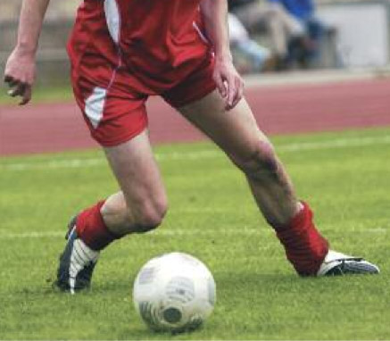

Studies clash over surgery’s significance
By Jordana Bieze Foster
In the wake of a Dutch report that surgery does not decrease the risk of radiographically diagnosed osteoarthritis following anterior cruciate ligament injury, research presented in July identified preoperative variables that could predict the development of OA after ACL reconstruction.
Researchers from Leiden University Medical Center in the Netherlands made headlines this summer with their findings that patients with ACL or meniscal injuries were no less likely to demonstrate radiographic signs of cartilage degeneration 10 years later if they had undergone ACL reconstruction or partial meniscectomy, respectively. That study was published in the August issue of Radiology.
The investigators followed up with 326 patients from a decade-old database of 855 patients, who were originally enrolled in a previous study comparing MRI findings to arthroscopic findings for subacute knee complaints.
The 48 patients with ACL ruptures 10 years earlier were 2.8 times more likely to have increased more than one point on the Kellgren-Lawrence scale at follow up, as well as 5.5 times more likely to have joint space narrowing in the medial tibiofemoral compartment and seven times more likely to have osteophytes in the medial femoral compartment. However, the odds ratios were not significantly different for the 16 patients who underwent ACL reconstruction than for those who were treated conservatively.
The authors theorized that the elevated risk associated with ACL rupture stems from an increase in internal tibial rotation and anterior tibial translation, which increases rotational pressure on the medial knee compartment and shear forces in the medial and lateral compartments.
In a study presented in July at the annual meeting of the American Orthopaedic Society of Sports Medicine, researchers from the University of Pittsburgh reported a 39% prevalence of radiographic knee OA in 249 patients who had undergone ACL reconstruction an average of 7.8 years earlier. Radiographic knee OA was defined as a difference in K-L score of at least two grades in one compartment or at least one grade in two or more compartments, compared to the contralateral limb.
The strongest predictors of knee OA after ACL reconstruction were length of follow up, body mass index, concurrent medial meniscectomy, and medial compartment chondrosis of grade 2 or higher at the time of surgery, according to Ryan Tianran Li, BSE, a medical student at the University of Pittsburgh School of Medicine, who presented the results at the AOSSM meeting.
Interestingly, the Dutch study did not find that a history of meniscectomy affected OA risk. In the 183 knees with meniscal tears, partial meniscectomy was performed in 129. Patients with meniscal tears had an elevated risk of increased K-L score; those with medial meniscal tears were more likely to develop joint space narrowing and osteophytes in the medial tibiofemoral compartment, whereas those with lateral meniscal tears had similar patterns of risk in the lateral tibiofemoral compartment. However, those odds ratios were not significantly different for patients who had undergone meniscectomy.
In a study published in the September 2008 issue of the American Journal of Sports Medicine, researchers from Lund University in Sweden found that of 22 patients who underwent ACL reconstruction, only those who also had a meniscal tear had gone on to develop radiographic tibiofemoral OA at follow up 15 years later.
ACL reconstruction has been thought to decrease the risk of knee OA because it addresses issues of laxity and instability that can be associated with conservatively treated ACL tears. More recently, the Lund University group analyzed 100 subjects whose ACL tears were initially treated conservatively and found that those with significant anterior sagittal knee laxity three months post-injury were more likely to have developed knee OA 15 years later. Those findings were e-published on July 29 by the Scandinavian Journal of Medicine & Science in Sports.
Australian study identifies stiffness as risk factor for hamstring injuries
An Australian study suggests that muscle stiffness contributes to the risk of hamstring injury in athletes, but raises questions as to whether effective interventions should focus on increasing or decreasing that stiffness.
Investigators from the University of Technology in Sydney prospectively assessed hamstring stiffness and leg stiffness in 136 professional Australian Rules football players prior to the start of their competitive season. During the season, 14 players suffered acute, noncontact hamstring injuries. Each injured player missed an average of more than three weeks of match play.
At baseline, hamstring stiffness was 11% higher and leg stiffness 5% higher in the players who would go on to suffer a hamstring injury than in players who were not injured. When individual limbs were analyzed, however, hamstring stiffness was significantly lower in the injured limb than the contralateral limb; leg stiffness was greater in the uninvolved limb. The findings were e-published on July 1 in the American Journal of Sports Medicine.
Excessive stiffness is thought to reflect a lack of flexibility, whereas insufficient stiffness is thought to reflect a lack of strength.
Balance testing could help diagnose acetabular labral tears noninvasively
Proprioceptive balance testing may be used to screen for acetabular labral tears in athletes with hip pain, according to research from Wake Forest University in Winston-Salem, NC.
The investigators used a force plate to measure center of pressure deviations during a static single leg postural sway test in eight patients with symptomatic hip labral tears and five matched control subjects. They found a significant difference between the symptomatic limb and the contralateral limb of the injured patients, and a trend toward significance between the injured limbs and the control subjects.
The findings suggest that center of pressure testing could help practitioners improve their diagnoses of labral tears, which can be difficult to confirm using other types of noninvasive testing.

“A lot of our decisions are made based on history and physical examination. We’re often struggling to make the diagnosis,” said Allston J. Stubbs, MD, an assistant professor of orthopaedic surgery at Wake Forest, who presented the results in July at the AOSSM meeting. “We now include dynamic balance assessment as part of our workup.”


