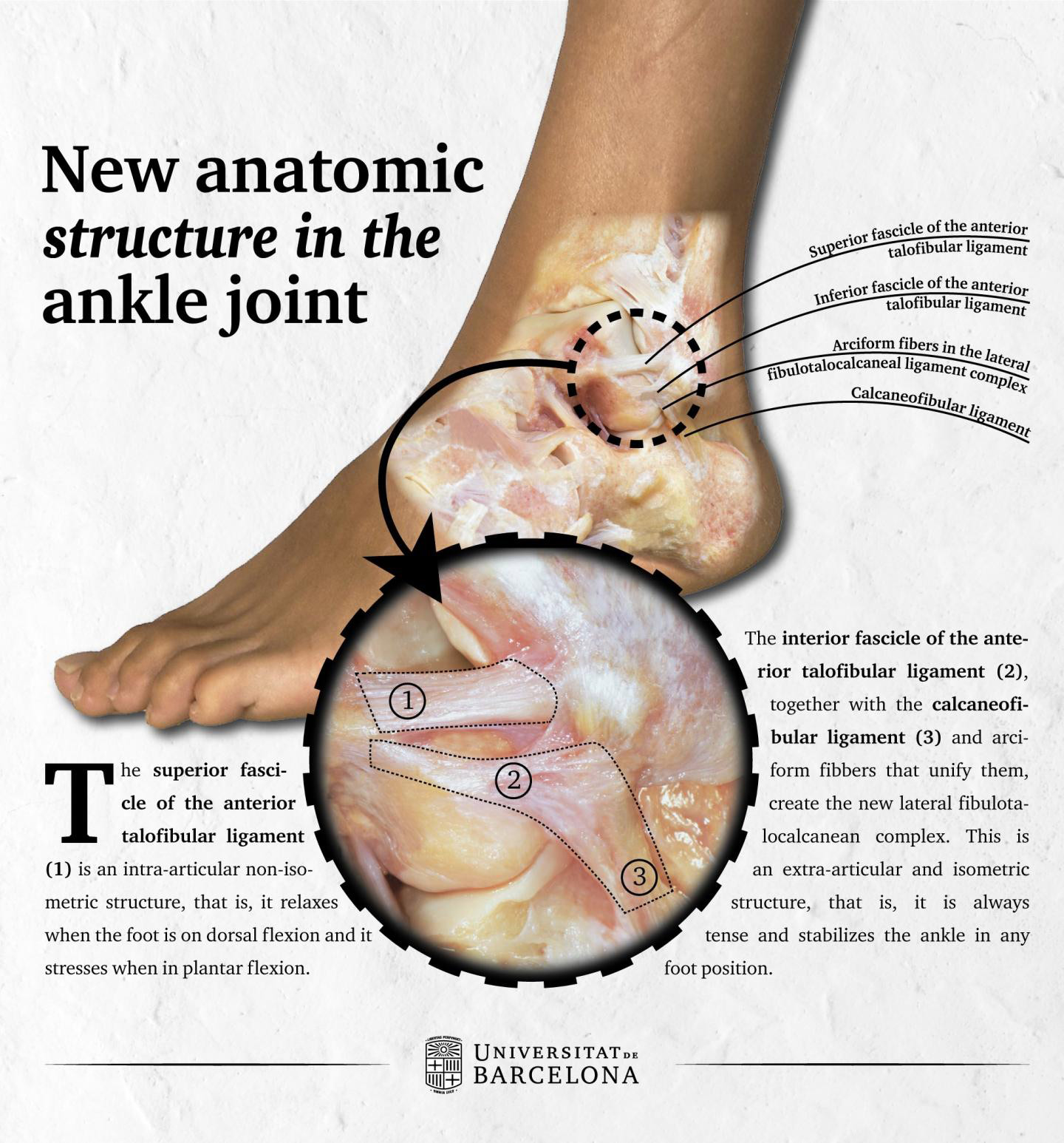
 Human anatomy guidelines group the ankle ligaments by two ligament complexes: the lateral collateral ligament and the medial or deltoid collateral ligament. Now, a University of Barcelona (UB) research team has defined a new anatomic structure in the ankle, the lateral fibulotalocalcaneal ligament complex (LFTCL). Also, for the first time, the researchers described one of the parts in this new structure as intra-articular. The results were published in the scientific journal Knee Surgery, Sports Traumatology, Arthroscopy. According to the researchers, this study changes the understanding of this joint and could explain why many ankle injuries produce chronic pain and lead to a propensity for further sprains, which have not yet been medically explained.
“This lack of explanation was the key to change the way to tackle ligament dissection in the dissection room and we saw linking fibers between ligaments that were usually removed because they were not associated with the ligament,” said Miquel Dalmau Pastor, PhD, PT, DPM, a researcher from the UB Human Embryology and Anatomy Unit and the UB Department of Pathology and Experimental Therapeutics.
According to the new study, these fibers link the inferior fascicle (set of ligamentous fibers) in the anterior talofibular ligament and the calcaneofibular ligament, two out of the three components in the lateral collateral ligament. “This connection was never described, and contrary to what was thought, it suggests that both ligaments are a functional unit,” said Pastor. “That is, we could consider these two connected ligaments as an anatomic structure we call lateral fibulotalocalcaneal ligament complex.”
This description of the ligament fits with some clinical publications that showed good results of isolated repairs of the anterior talofibular ligament in cases of injuries of the anterior talofibular ligament and calcaneofibular ligament. The careful dissection of the articular capsule in the ankle enabled researchers to identify the intra-articular compound in the anterior talofibular ligament. According to the study, this ligament would be built by two fascicles, the superior and inferior one, where the superior fascicle lies within the joint and the inferior one is outside. This inferior fascicle together with the calcaneofibular ligament and fibers would form the fibulotalocalcaneal lateral complex, which would then be an extra-articular structure.
“These findings suggest the behavior after an injury will be similar to the other intra-articular ligaments, such as the twill, which are not able to cicatrize, and this makes the joint remain unstable and in many cases it requires a surgical operation,” said Pastor. These results would explain why many sprains cause pain even after the patient follows physician-prescribed treatment.
Source: Vega J; Malagelada F; Manzanares MC; Dalmau Pastor M. The lateral fibulotalocalcaneal ligament complex: An ankle stabilizing isometric structure. Knee Surgery, Sports Traumatology, Arthroscopy, 2018. Doi: 10.1007/s00167-018-5188-8.


