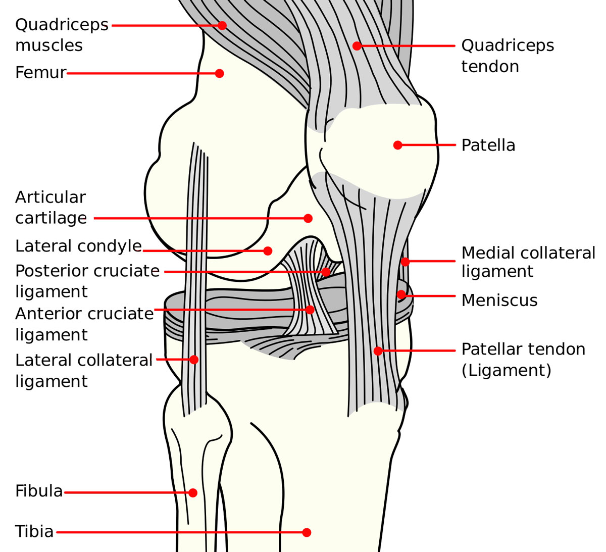
Right knee, seen from an angle between anteriorly and laterally.
Osteoarthritis (OA) affects nearly 50% of patients who undergo anterior cruciate ligament reconstruction (ACLR) within 2 decades after surgery. The mechanism of OA development has been elusive; many experts theorize that disruption of knee–joint tissue homeostasis is key. Now, researchers, using ultrasonography (US), have found an increase in the thickness of the anterior femoral cartilage and cross-sectional area 3 years postoperatively.
The multidisciplinary team of researchers—from Tufts Medical Center, University of Massachusetts Medical School, University of North Carolina at Chapel Hill, University of Tennessee, and University of Glasgow—used US to examine the anterior femoral cartilage cross-sectional area and thickness in both injured and contralateral limbs of study participants, as well as in the limbs of uninjured healthy volunteers.
In their case–control study of 20 participants who had undergone ACLR, compared to 28 uninjured controls, researchers found that the mean thickness of the greater anterior femoral cartilage cross-sectional area was 96.68 mm2 (±26.68 mm2) in the injured limb, compared to 85.69 mm2 (±17.57 mm2) (P<.001) in the contralateral limb and to 84.62 mm2 (±15.89 mm2) (P=.04) in uninjured controls. To assess the cross-sectional area, researchers first outlined the femoral cartilage on each US image; femoral cartilage thickness was then measured as the length of the straight line drawn from the cartilage–bone interface to the synovial space–cartilage interface.
More specifically, mean greater medial condyle thickness of 2.61 mm (±0.61 mm) was seen in the injured limbs, compared to contralateral limbs, at 2.36 mm (±0.47 mm) (P=.01), and to uninjured limbs, at 2.22 mm (±0.40 mm) (P=.01). Greater lateral condyle thickness in injured limbs had a mean of 2.46 mm (±0.65 mm), compared to 2.12 mm (±0.41 mm) in uninjured limbs (P=.03).
On average, these changes were evident 37 months after surgery. Researchers noted that, the further out from ACLR, the greater the femoral cartilage area; however, time was not associated with thickness of the medial condylar, intercondylar, or lateral condylar femoral cartilage.
This study builds on the work of other investigators, using magnetic resonance imaging, which revealed worsening cartilage composition as early as 1 year after ACLR. When viewed together, the aggregate data suggest that progressive thickening of cartilage after ACLR is detectable using US—an imaging modality that is accessible and inexpensive. Future studies, the researchers said, should assess ACLR patients at longer intervals to understand when cartilage thinning, which can lead to OA, actually begins.
Source: Harkey MS, Blackburn JT, Nissman D, et al. Ultrasonographic assessment of femoral cartilage in individuals with anterior cruciate ligament reconstruction: a case-control study. J Athl Train. 2018;53(11):1082-1088.


