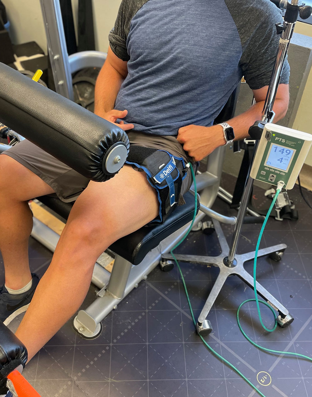

Knee-extension exercise performed with blood flow restriction. The tourniquet is placed above the active muscles and inflated to a pressure that is controlled automatically through a Personalized Tourniquet System. Here, a low-load (20% of the volunteer’s 1-repetition maximum) is applied with the tourniquet pressure set to 60% of the lowest effective occlusion pressure.
By Chris Pignanelli, PhD(c)
With benefits similar to traditional training practices but with less work, BFR exercise is gaining ground in rehabilitation settings as well as athletic training rooms.
The intentional manipulation of blood flow during exercise, commonly referred to as blood flow restriction (BFR) exercise, has piqued the interest of scientists and practitioners alike over the past two decades. The aim of BFR exercise is to reduce the arterial blood supplying the active limb muscles and simultaneously prevent venous blood from leaving them. This reduction in blood supply can be accomplished by applying external pressure with a tourniquet above the contracting muscles set to a percentage (~40% – 100%) of an individual’s lowest effective occlusion pressure (also referred to as arterial occlusion pressure). The lowest effective occlusion pressure can be determined automatically by custom tourniquet systems (eg, Delfi Medical; Vancouver, BC, Canada) or manually by measuring the tourniquet pressure required to cause the disappearance of a distal artery pulse. While other methods to elicit BFR have been used, such as elastic wraps or straps, caution is warranted as it is difficult to know the pressures that are applied with these techniques. BFR can be continuously applied throughout both the exercise sets and rest periods, or intermittently during only the exercise sets or rest periods.
Why is BFR of interest to practitioners?
The application of BFR is probably best known for its use to increase muscular size and strength during ‘low-load’ resistance exercise (less than 40% of an individual’s 1-repetition maximum). Indeed, the addition of BFR might promote similar adaptations to traditional resistance exercises but with less work and lower mechanical loading, which may have implications for its prescription to certain populations. BFR can also be used during movements that have traditionally not been thought to improve muscular function and/or overall fitness. For example, low-intensity treadmill walking and cycling with BFR can increase muscular size and strength or cardiorespiratory fitness (inferred through pulmonary maximal oxygen uptake; V̇O2max).1-4 Furthermore, BFR has been successfully applied during low-moderate intensity interval aerobic exercise5,6 and even with transcutaneous electrical muscle stimulation (TEMS).7 Such diversity has led to many potential applications of BFR in both a rehabilitation setting and to maximize training adaptations in athletic populations, which is covered in more detail below.
Application of BFR to attenuate muscle disuse
During muscle disuse, such as with injury or bed rest, a rapid loss in muscle function occurs. For instance, it has been estimated that as little as 2 days of limb immobilization causes an approximate 2% reduction in quadriceps muscle size and by 7 days, approximately 6.7%.8 This muscle atrophy coincides with an approximate 20% decrease in dynamic force production. In comparison, 84 days of strict bed rest leads to an average 17% reduction in quadriceps muscle size and 36% – -43% reduction in maximal quadriceps force production.9 Together, these findings suggest almost half of the muscle atrophy and loss in strength can occur within the first week and emphasize the importance of early interventions to combat these negative outcomes.
The addition of BFR exercise to traditional rehabilitation practices may offer an effective means to help attenuate the early detrimental effects of muscle disuse. This is in part due to lower mechanical loading required to achieve beneficial effects of exercise. Indeed, low-load resistance exercise with BFR was an effective alternative to traditional high-load resistance exercise to regain muscle size and strength during an 8-week ACL reconstruction rehabilitation program.10 Further, the authors also demonstrated that the addition of BFR exercise improved several clinical outcomes such as range of motion, knee swelling, and perceived pain to a greater extent than the traditional resistance exercise program. While a promising finding, the application of resistance exercise (regardless of BFR) may not be feasible in some cases, such as when complete limb unloading is required, or in patients with limited range of motion, impaired ability to voluntarily activate muscles, and/or equipment or space limitations. As such, innovative solutions are required to circumvent these limitations. This may include the application of BFR without exercise. However, while some evidence suggests applying BFR in a cyclical manner without muscle contractions can attenuate muscle atrophy and/or strength loss,11-13 this has not been universally reported.14
Seeing how BFR with low-intensity transcutaneous electrical muscle stimulation (TEMS) may be superior to low-intensity TEMS alone at augmenting muscle size and strength in healthy individuals,7 our lab investigated whether BFR and TEMS could effectively attenuate muscle atrophy and strength loss during muscle disuse.15 Participants were divided into one of 3 groups during a 2-week limb immobilization protocol: control group with no intervention, BFR only (2x daily, 5 days/week), or BFR and TEMS (2x daily, 5 days/week). Interestingly, only BFR with TEMS prevented muscle atrophy compared to the control group, whereas BFR alone did not. Despite the attenuation in muscle atrophy, maximal force production was not preserved with BFR and TEMS. While these findings are promising, future work is required to compare the efficacy of BFR with TEMS to TEMS alone in preventing muscle atrophy and their potential to improve clinical outcomes in rehabilitation populations.
Application of BFR to improve the physical preparation in athletes

Treadmill walking performed with blood flow restriction. The tourniquet is placed above as proximal as possible on the legs and inflated to a pressure that is controlled automatically through a Personalized Tourniquet System. Treadmill walking was performed at a 5% incline and low-speed (5 km/hr) with the tourniquet pressure set to 100% of the lowest effective occlusion pressure.
Most of the early research focused on BFR exercise in untrained populations with no previous or limited training experience. While these studies provided important information, it remained unclear whether well-trained athletes could also benefit from BFR exercise. Luckily, several investigations over the past decade have examined whether BFR exercise could potentiate training adaptations in well-trained athletes. For example, whole-muscle quadriceps size and type I muscle fibers (considered the ‘oxidative’ fibers) increased by 3% – 12% from baseline following a 6.5-week training block that added only 10 sessions of front squats with BFR in national-level powerlifters, whereas no changes in muscle size occurred in powerlifters who trained without BFR.16 These findings suggest the addition of BFR exercise could be an effective method to augment muscle size in a short time frame in well-trained athletes. On the opposite end of the athlete ‘spectrum’, endurance-trained athletes may also benefit from the addition of BFR into their regular training. The application of BFR during low-intensity rowing 17 and in the rest periods of bike sprints18-19 have both been shown to improve V̇O2max (~4% – 9%) and maximal aerobic power output (~4% – 15%) in already well-trained endurance athletes in as little as 2-3 sessions/week for 4 – 5 weeks. While the underlying mechanisms of this improved V̇O2max and direct sport performance benefits are unclear, these results suggest that BFR is also a promising strategy to potentiate aerobic training adaptations.
Conclusion
The diversity of settings for which BFR can be applied has led to great interest in many different scenarios ranging from rehabilitation to the physical preparation of athletes. While this diversity can be viewed as a positive, it could also make it challenging for practitioners to successfully apply this technique. To circumvent this issue, a position statement by Patterson et al20 provides general guidelines for BFR use, such as the applied pressures, duration, and sets with different BFR exercise modalities.
In total, the studies discussed above provide proof of principle evidence for the use of BFR to positively impact both rehabilitation and athletic training practices. However, it is worth noting this does not suggest BFR exercise replaces traditional practices. Instead, BFR may act as an effective adjunct modality that is incorporated with traditional rehabilitation methods or specific training blocks where certain physiological adaptations are desired.
Chris Pignanelli is a PhD candidate in the Department of Human Health and Nutritional Sciences at the University of Guelph in Ontario, Canada. He can be reached at cp******@******ph.ca
BRF in Chronic Ankle Instability
A recent study published in the Journal of Sport Rehabilitation looked at the effects of BFR on lower-extremity muscle activation during dynamic balance exercises in individuals with chronic ankle instability. Participants did dynamic balance reaching exercises under normal/control conditions and BFR with a cuff around the thigh. Using electromyography, the researchers recorded greater muscle activation of the vastus lateralis and soleus during the BFR sets, with no differences seen in the tibialis anterior or the fibularis longus between conditions. The authors observed greater ratings of perceived postural instability and exertion during balance exercises with BFR than control.
Read more at Burkhardt M, et al. Effects of blood flow restriction on muscle activation during dynamic balance exercises in individuals with chronic ankle instability. J Sport Rehab. 2021;30(6):870-875.
- Abe T, Kearns CF, Sato Y. Muscle size and strength are increased following walk training with restricted venous blood flow from the leg muscle, Kaatsu-walk training. J Appl Physiol. 2006;100:1460–1466.
- Abe T, Sakamaki M, Fujita S, et al. Effects of low-intensity walk training with restricted leg blood flow on muscle strength and aerobic capacity in older adults. J Geriatr Phys Ther. 2010(b); 33: 34–40.
- Abe T, Fujita S, Nakajima T, et al. Effects of low-intensity cycle training with restricted leg blood flow on thigh muscle volume and VO2max in young men. J Sports Sci Med. (2010a);9(3):452-458.
- Park S, Kim JK, Choi HM, Kim HG, Beekley MD, Nho H. Increase in maximal oxygen uptake following 2-week walk training with blood flow occlusion in athletes. Eur J Appl Physiol. (2010); 109(4):591–600.
- Corvino RB, Oliveira MFM, Denadai BS, Rossiter HB, Caputo F. Speeding of oxygen uptake kinetics is not different following low-intensity blood-flow-restricted and high-intensity interval training. Exp Physiol. 2019;104(12):1858–1867.
- Christiansen D, Eibye K, Hostrup M, Bangsbo J. Training with blood flow restriction increases femoral artery diameter and thigh oxygen delivery during knee-extensor exercise in recreationally trained men. J Physiol. 2020;598(12):2337–2353.
- Natsume T, Ozaki H, Saito AI, Abe T, Naito H. Effects of electrostimulation with blood flow restriction on muscle size and strength. Med Sci Sports Exerc. 2015;47(12):2621–2627.
- Kilroe SP, Fulford J, Jackman SR, van Loon LJC, Wall BT. Temporal muscle-specific disuse atrophy during one week of leg immobilization. Med Sci Sport Exerc. 2020;52(4):944-954.
- Trappe S, Trappe T, Gallagher P, Harber M, Alkner B, Tesch P. Human single muscle fibre function with 84 day bed-rest and resistance exercise. J Physiol. 2004;557(Pt 2):501–513.
- Hughes L, Rosenblatt B, Haddad F, et al. Comparing the effectiveness of blood flow restriction and traditional heavy load resistance training in the post-surgery rehabilitation of anterior cruciate ligament reconstruction patients: a UK National Health Service randomised controlled trial. Sports Med. 2019;49(11):1787–1805.
- Takarada Y, Takazawa H, Ishii N. Applications of vascular occlusion diminish disuse atrophy. Med Sci Sports Exerc. 2000;32(12):2035–2039.
- Kubota A, Sakuraba K, Koh S, Ogura Y, Tamura Y. Blood flow restriction by low compressive force prevents disuse muscular weakness. J Sci Med Sport. 2011;14(2):95–99.
- Barbalho M, Rocha AC, Seus TL, Raiol R, Del Vecchio FB, Coswig VS. Addition of blood flow restriction to passive mobilization reduces the rate of muscle wasting in elderly patients in the intensive care unit: a within-patient randomized trial. Clin Rehabil. 2019;33(2):233–240.
- Iversen E, Røstad V, Larmo A. Intermittent blood flow restriction does not reduce atrophy following anterior cruciate ligament reconstruction. J Sport Health Sci. 2016;5(1):115–118.
- Slysz JT, Boston M, King R, Pignanelli C, Power GA, Burr JF. Blood flow restriction combined with electrical stimulation attenuates thigh muscle disuse atrophy. Med Sci Sports Exerc. 2021;53(5):1033–1040.
- Bjørnsen T, Wernbom M, Kirketeig A, et al. Type 1 muscle fiber hypertrophy after blood flow-restricted training in powerlifters. Med Sci Sports Exerc. 2019;51(5):288–298.
- Held S, Behringer M, Donath L. Low intensity rowing with blood flow restriction over 5-weeks increases V̇O2max in elite rowers: A randomized controlled trial. J Sci Med Sport. 2020;23(3):304–308.
- Taylor CW, Ingham SA, Ferguson RA. Acute and chronic effect of sprint interval training combined with postexercise blood-flow restriction in trained individuals. Exp Physiol. 2016;101(1):143–154.
- Mitchell EA, Martin NRW, Turner MC, Taylor CW, Ferguson RA. The combined effect of sprint interval training and postexercise blood flow restriction on critical power, capillary growth , and mitochondrial proteins in trained cyclists. J Appl Physiol. 2019;126(1):51–59.
- Patterson SD, Hughes L, Warmington S, et al. Blood flow restriction exercise: considerations of methodology, application, and safety. Front Physiol. 2019;10:533.


