 The use of detailed orthopedic imaging to examine dancers’ ankles while in the en pointe position offers insight into the biomechanical demands associated with a position that, although highly unnatural, is nevertheless essential to a ballerina’s performance.
By Jeffrey A. Russell, PhD, AT, FIADMS
The beauty and grace of ballet en pointe disguises an impressive forced arrangement of the bones of the ankle. A look deep down inside reveals a fascinating picture—one might call it a marvel of adaptive anatomy. Looking closely at the actual positions of the ankle bones, one must acknowledge with respect the striking ability ballet dancers possess to stand and dance en pointe.
Dancing en pointe (on the tips of the toes) is not for the faint of heart, nor for the faint of ankles. It is a rigorous subdiscipline of ballet, and one that can be fraught with difficulties and unsafe practices that may lead to foot and ankle injuries in immature or weak tissues. One frequent problem is the practice of allowing young girls to dance en pointe at a younger age than is advisable and without the necessary skeletal, muscular, and technical development.
Unfortunately, age by itself is not an acceptable criterion with which to make decisions about starting pointe work. This is one reason that authors have identified appropriate guidelines for beginning pointe training,1,2 and why the International Association for Dance Medicine & Science provides a resource sheet (available in several languages) to assist dancers, dance teachers, and dance parents with making wise decisions about initiating pointe.3

Figure 1: X-ray of a ballet dancer showing the bony anatomy of the foot and ankle.
Only one study is known to have investigated injuries related to dancing en pointe specifically;4 it was preliminary and had several limitations. However, research has shown repeatedly that a substantial number of injuries occurring in professional ballet—in some reports a majority—occur to the foot and ankle.5-10 Therefore, the presumption can be made that, because pointe work is integral to the repertoire of most professional classical ballet companies, it is likely that dancing en pointe contributes to this injury rate because of the extreme foot and ankle positioning it requires.11
A research window into the relative positions of the bones of the ankle and foot has been opened using orthopedic imaging.12-15 These studies show precise orientations of the bones, using radiography12,13 and magnetic resonance imaging (MRI),14,15 and of the soft tissues, using MRI.14,15 When viewed with these modalities, it is clear the en pointe position is not natural—and is likely injurious. However, with pointe work so important to the ballet aesthetic, it serves the dance and dance medicine communities well to understand the demands of this position with a view to ensuring that ballet dancers, particularly young ones whose teachers and parents are contemplating starting them en pointe, are as safe as possible in the genre.
Anatomy and stability

Figure 2: X-ray of a ballet dancer en pointe. The black arrow points to the area where the tibia, talus, and calcaneus converge when a dancer is en pointe. The black dotted line identifies the posterior portion of the talus beyond its articular surface. The white dotted line denotes the corresponding portion of the tibia’s articular surface that rests against the posterior talus in this position.
The standing x-ray of a ballet dancer in Figure 1 identifies the bones of the leg, ankle, and foot. The talocrural joint comprises the distal portions of the tibia and fibula (which form a box-like mortise) and the talus. The rounded upper part, or dome, of the talus fits into the mortise; in typical standing, primary weightbearing in this joint occurs in the articulation of the distal tibia’s hyaline cartilage surface and the hyaline cartilage of the talar dome. However, in an en pointe stance, weightbearing of the tibia moves progressively posterior on the talar dome and, in fact, the posterior portion of the tibia’s articular surface leaves the articular surface of the dome to rest on the posterior talus15 as the posterior tibia, posterior talus, and superior calcaneus converge.14 The en pointe x-ray in Figure 2 shows this positioning.
The convergence of the three bones is significant because this “locks” the ankle into a more stable position en pointe16,17 than might otherwise be predicted, based on the fact that the narrowest aspect of the talar dome—the posterior aspect—is placed into the ankle mortise during plantar flexion.18 That is to say, with the narrow posterior dome in the mortise, one might expect relative joint laxity,19 but the locking function overrides this. Furthermore, MRI shows more clearly than x-rays how the positioning of the tibia’s articular surface is partially incongruent with the joint surface of the talus (Figure 3). This may also provide stability, thanks to the roughness of the nonarticular joint surface of the posterior talus resting under the tibial plafond during full weightbearing en pointe.
Parenthetically, in spite of the extreme plantar flexion seen in the talocrural joint, the position of the foot during pointe dancing cannot rely on this motion alone. Attaining the full en pointe position requires contributions from movements between the bones in the foot.12,20,21 Examples of such movements include sliding between the talus and the navicular, the navicular and the medial cuneiform, and the medial cuneiform and the first metatarsal. These small increments of motion combine to provide approximately 30% of the plantar flexion range.12 In addition, it is noteworthy that the talus shifts slightly anterior from under the tibia as the ankle-foot complex moves en pointe.15 This subtlety arises because the converging tibia, talus, and calcaneus form a fulcrum that applies an anterior force to the talus, somewhat like a watermelon seed being squeezed from between one’s thumb and forefinger.
Utility of MRI in visualizing en pointe
MRI is an ideal method of viewing the tissues of the foot and ankle because, unlike x-rays, it provides a visual depiction of multiple types of structures, not just bones. In addition, MR imaging does not expose patients to ionizing radiation. However, imaging the ankle and foot en pointe presents special challenges, and there is one exception to the utility of MRI for this application: the inability to incorporate a high-field strength scanner (ie, one with a strong magnet that can produce very detailed images) for imaging a dancer in the functional weightbearing en pointe position.

Figure 3: Proton density-weighted magnetic resonance image of the ankle en pointe with spectral attenuated inversion recovery (SPAIR) fat suppression taken by a 3T MR scanner. This dancer was supine with her foot, ankle, and leg bound in the en pointe position according to the method described in the article. The circle shows the area of tibia, talus, and calcaneus convergence. The white dotted line indicates the portion of the tibia’s articular surface that lies against the nonarticular posterior talus.
Prior to the work presented in this article, MR imaging was used to assess potential ankle conditions in dancers.9,11,22 The goal of these studies was primarily the identification of anatomical variants, such as a protruding posterior malleolus of the tibia or superior tubercle of the calcaneus, or the assessment of bone edema. However, more fundamental than these variants is the functional anatomy of the ankle. This is because an understanding of how the components of the ankle and foot align forms the framework for teachers and practitioners for helping dancers be as healthy as possible when they undertake pointe training.
“Open” MR scanners exist that allow an individual to be upright and weightbearing within a scanner instead of in the typical supine position; these have been used previously to scan dancers standing en pointe (Figure 4).14,23 However, the magnets used in these studies are relatively weak (.25-.30 Tesla). Open scanners also are limited in this application by the short amount of time a dancer can remain en pointe, especially motionless. In addition to the field strength limiting the quality of the images, the scanning parameters must be set suboptimally for the short scans, and any wavering by a dancer as she balances on her toes creates artifacts on the images that can render them difficult, if not impossible, to read accurately.
Incidentally, a technical point of this method is that pointe shoes without sole nails are required, as ferromagnetic nails disrupt the magnetic field, creating unusable MR images (not to mention a safety hazard). Gaynor Minden is one company manufacturing shoes that do not contain nails in their soles; these were selected for use in the studies described herein.
To mitigate the problems associated with open MRI units, we developed a supine “en pointe” protocol.15 Replicating as true an en pointe position as possible requires a dancer to wear a pointe shoe and stand en pointe while her foot, ankle, and leg are splinted with wood rods wrapped with Velcro straps (or other nonferromagnetic materials). Once affixed this way, the dancer is placed on her back in a standard high-field strength (3 Tesla [3T]) MR scanner with her ankle inside a knee coil, which helps focus the radiofrequency signals to optimize the resulting images (Figure 5). A similar protocol, with the coil repositioned and the MRI parameters adjusted, could be used to visualize the foot in an en pointe position.
In one study15 we used this MRI protocol to review uninjured ankles in six university-level dancers who had been dancing for an average of 13 years and dancing en pointe for an average of seven years. The dancers typically reported minor amounts of dancing-related pain in their ankles and feet. All exhibited several traits in their ankle MRIs: the posterior portion of the articular surface of the tibia rested on a nonarticular surface of the posterior talus; the synovial sheaths of the flexor and fibularis tendons collected fluid; Kager’s fat pad was impinged by the posterior tibial plafond; and small ganglion cysts were apparent in one or more spots around the ankle and proximal foot.
The images obtained with high-field strength MRI (Figure 3) are of substantially higher quality than those obtained with the low-field strength standing open MR procedure. It appears from the images taken with this adapted method that the en pointe position is essentially preserved even though the dancers are back-lying rather than weightbearing. There are no qualitative differences in the positions of the bones when evaluated on the images from both techniques, and the finding that the tibia, talus, and calcaneus converge in every case holds true when the dancers are imaged supine15 as it has in previous studies when they have been imaged while standing en pointe in an open MR device.14
Ongoing research
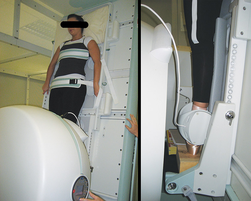
Figure 4: A dancer standing in an open MR scanner. The right image shows of her lower extremity with the ankle in a knee coil.
X-ray and MR imaging are useful for evaluating the extreme position of the ankle and foot required to attain the en pointe position in ballet. The observation that the talocrural joint exhibits incongruence when a dancer is en pointe leads to a logical next question: Does the fact that the articular surface of the talus rests on the nonarticular posterior talus increase the likelihood of ballet dancers developing early ankle osteochondritis or osteoarthritis?

Although limited research on these in ballet dancers is published, available reports are equivocal about the presence of degeneration in the ankle.24-26 Thus, an important development in MR imaging may be helpful for more accurately discerning whether ankle osteoarthritis occurs in dancers’ ankles. Mapping of T2/T1ρ MRI signals27,28 allows closer investigation of hyaline cartilage quality than is possible with traditional MR imaging; it analyzes the relative presence of proteoglycans and other important molecules in the cartilage’s extracellular matrix. Hopefully, future research using this technique will shed additional light on the extent to which pointe dancing affects the ankle’s joint surfaces.

Figure 5: A dancer positioned supine, with her foot, ankle, and leg splinted, for entry into a Philips 3T MR scanner.
The supine method of high-field strength MR imaging of ballet dancers’ ankles in the en pointe position is helpful clinically: Because the ankle’s structures can be viewed in the relative positions in which they interact during pointe dancing it identifies structures and pathologies that can hinder this type of dancing. Although routine MR imaging of all young dancers wanting to dance en pointe is neither feasible nor advised, possible further applications of this method in select dancers could include pre-emptively evaluating positions of the bones or soft tissues that could lead to injury, with the goal of initiating preventive conditioning or retraining. Additionally, the technique may allow surgeons contemplating surgical intervention in pointe dancers (eg, for os trigonum removal) to better visualize both the pathology and the intended operative results.
Conclusion
Dancing en pointe is a beautiful and essential aspect of ballet that has been entrenched since Marie Taglioni’s progressive foray in La Sylphide during the 1830s. It is the dream of young girls who take ballet lessons to obtain their first pointe shoes and begin training in this type of dance. However, such training is not without a physical cost that must be understood and ameliorated to the greatest possible degree within the context of the aesthetics of ballet.
The use of orthopedic imaging to examine pointe dancers’ ankles in detail offers insight into the demands placed on the ankle by dancing en pointe, as these radiographic methods allow assessment of the bones’ alignment. In short, this most unnatural of ankle positions causes incongruence of the proximal (on the tibia) and distal (on the talus) articular surfaces of the ankle joint such that a section of the hyaline cartilage of the tibia rests on a nonarticular portion of the talus.
With pointe dancing here to stay in ballet, these research findings suggest that dance teachers, dance medicine clinicians, and others responsible for the healthy practice of dance must help dancers achieve their performance goals in a safe way. This includes moderating young dancers’ commencement of pointe work until they are maturationally and physically (and emotionally, for that matter) ready to take up this high-level training, a worthy goal suggested by leading clinicians and dance medicine organizations.
Jeffrey A. Russell, PhD, AT, FIADMS, is assistant professor of athletic training and director of Science and Health in Artistic Performance at Ohio University in Athens. He is a fellow of the International Association for Dance Medicine & Science.
1. Shah S. Determining a young dancer’s readiness for dancing on pointe. Curr Sports Med Rep 2009;8(6):295-299.
2. Weiss DS, Rist RA, Grossman G. When can I start pointe work? Guidelines for initiating pointe training. J Dance Med Sci 2009;13(3):90-92.
3. International Association for Dance Medicine & Science. Guidelines for Initiating Pointe Training. 2016 IADMS website. http://www.iadms.org/?page=pointe. Accessed June 6, 2016.
4. Nunes NMA, Haddad JJ, Bartlett DJ, Obright KD. Musculoskeletal injuries among young, recreational, female dancers before and after dancing in pointe shoes. Pediatr Phys Ther 2002;14(2):100-106.
5. Allen N, Nevill A, Brooks J, et al. Ballet injuries: injury incidence and severity over 1 year. J Orthop Sports Phys Ther 2012;42(9):781-790.
6. Allen N, Nevill AM, Brooks JHM, et al. The effect of a comprehensive injury audit program on injury incidence in ballet: a 3-year prospective study. Clin J Sport Med 2013;23(5):373-378.
7. Arendt YD, Kerschbaumer F. [Injury and overuse pattern in professional ballet dancers]. Z Orthop Ihre Grenzgeb 2003;141(3):349-356.
8. Byhring S, Bø K. Musculoskeletal injuries in the Norwegian National Ballet: a prospective cohort study. Scand J Med Sci Sports 2002;12(6):365-370.
9. Hillier JC, Peace K, Hulme A, Healy JC. MRI features of foot and ankle injuries in ballet dancers. Br J Radiol 2004;77:532-537.
10. Nilsson C, Leanderson J, Wykman A, Strender L. The injury panorama in a Swedish professional ballet company. Knee Surg Sports Traumatol Arthrosc 2001;9(4):242-246.
11. Elias I, Zoga AC, Raikin SM, et al. Bone stress injury of the ankle in professional ballet dancers seen on MRI. BMC Musculoskelet Disord 2008;9:39.
12. Russell JA, Shave RM, Kruse DW, et al. Ankle and foot contributions to extreme plantar flexion and dorsiflexion in female ballet dancers. Foot Ankle Int 2011;32(2):183-188.
13. Russell JA, Shave RM, Kruse DW, et al. Is goniometry suitable for measuring ankle range of motion in female ballet dancers? An initial comparison with radiographic measurement. Foot Ankle Spec 2011;4(3):151-156.
14. Russell JA, Shave RM, Yoshioka H, et al. Magnetic resonance imaging of the ankle in female ballet dancers en pointe. Acta Radiol 2010;51(6):655-661.
15. Russell JA, Yoshioka H. Assessment of female ballet dancers’ ankles in the en pointe position using high field strength magnetic resonance imaging. Acta Radiol 2015 Nov 13. [Epub ahead of print]
16. Hamilton WG. Sprained ankles in ballet dancers. Foot Ankle 1982;3(2):99-102.
17. Shah S, Luftman J, Vigil DV. Stress injury of the talar dome and body in a ballerina: a case report. J Dance Med Sci 2005;9(3):91-95.
18. Sammarco GJ, Tablante EB. Foot and ankle in dance. In: Sataloff RT, Brandfonbrener AG, Lederman RJ, eds. Performing Arts Medicine. 2nd ed. San Diego: Singular Publishing Group; 1998: 301-320.
19. Brown TD, Micheli LJ. Foot and ankle injuries in dance. Am J Orthop 2004;33(6):303-309.
20. Hamilton WG, Hamilton LH, Marshall P, Molnar M. A profile of the musculoskeletal characteristics of elite professional ballet dancers. Am J Sports Med 1992;20(3):267-273.
21. Novella TM. Simple techniques for quantifying choreographically essential foot and ankle extents of motion. J Dance Med Sci 2004;8(4):118-122.
22. Peace KA, Hillier JC, Hulme A, Healy JC. MRI features of posterior ankle impingement syndrome in ballet dancers: a review of 25 cases. Clin Radiol 2004;59(11):1025-1033.
23. Graber OP, Oberthaler W. [Weight bearing upright magnetic resonance imaging of pointe dancing]. Röntgenpraxis 2008;56(5):195-198.
24. Andersson S, Nilsson B, Hessel T, et al. Degenerative joint disease in ballet dancers. Clin Orthop 1989;238:233-236.
25. Angioi M, Maffulli G, Morrissey D, et al. Early signs of osteoarthritis in professional ballet dancers: A preliminary study. Clin J Sport Med 2014;24(5):435-437.
26. van Dijk CN, Lim LSL, Poortman A, et al. Degenerative joint disease in female ballet dancers. Am J Sports Med 1995;23(3):295-300.
27. Menezes NM, Gray ML, Hartke JR, Burstein D. T2 and T1rho MRI in articular cartilage systems. Magn Reson Med 2004;51(3):503-509.
28. Wang L, Regatte RR. T1rho MRI of human musculoskeletal system. J Magn Reson Imaging 2015;41(3):586-600.


