
Photo courtesy of Ossur Americas
Dose-response research refutes the common perception that increasing brace wear time leads to muscle atrophy in patients with knee osteoarthritis. In fact, longer bracing duration appears to improve hamstring strength as well as increasing patients’ physical activity levels.
By Cheryl L. Hubley-Kozey, PhD, and Gillian Hatfield Murdock, PT, MSc
Osteoarthritis (OA), the most prevalent form of arthritis,1 is characterized by the breakdown of articular cartilage. This breakdown results from an imbalance between cartilage synthesis and degradation triggered by biochemical and biomechanical processes.2 More than 27 million individuals in the US3 and 4.4 million Canadians1 report having OA. Given the aging population, increased obesity rates, and sedentary lifestyles, it’s estimated that 25% to 30% of the North American population will develop OA within the next 20 to 30 years.1,3
While articular cartilage changes are the hallmark of OA, other joint structures including bone, ligaments, muscles, and nerves are also affected, leading to pain and physical function limitations.4 These functional limitations contribute to the staggering economic burden of OA. In the last decade, direct health care expenditures reached $328 billion5 and more than $10 billion1 in the US and Canada, respectively.
The knee is the joint most often affected by OA. For adults older than 65, knee OA results in more difficulty with typical activities of daily living, such as walking and climbing stairs, than any other medical condition.6 However, what is more alarming is that the group of adults aged 45 to 54 years is growing more rapidly than any other age group with knee OA.7
There is no cure for knee OA and total knee arthroplasty is the end-stage treatment for advanced disease. Joint arthroplasty is not a practical solution for those in younger age categories as implants have a lifespan of 15 to 20 years, and patient satisfaction and outcomes dramatically decrease following revision surgery.8 Researchers once thought OA was primarily a degenerative process, but the thinking over the past decade has changed to support the idea that OA is a metabolically active process that can be delayed with an optimal environment.4 Hence, there is an urgent need to develop interventions that delay OA progression.9
Pharmaceutical agents have been the mainstay of OA management, particularly the management of OA-related pain. Although pain relief has a positive impact on quality of life, long-term pain medication usage has been associated with an increased risk of tibial cartilage defects10 and knee OA progression.11 It is thought that the increased risks could be due to deleterious effects on the articular cartilage or to excessive loading on an analgesic-affected joint.11 The latter hypothesis is supported by reports of increased joint loading following pain relief in those with moderate knee OA.12,13 For these reasons, international knee OA management guidelines recommend that the initial OA treatment should include nonpharmacological interventions and, if pharmacological pain-relieving agents are used, they should not be used long-term.14,15
Disease-modifying pharmacological agents are also being developed to disrupt the imbalanced biochemical processes associated with OA16 along with biological products to replace damaged tissue,17 but there is also a focus on altering the biomechanical environment. Based on evidence from animal models,18-20 theoretical models,21,22 and empirical data,23 loading forces are considered catalysts for initiating the degenerative process and for disease progression. The theory is that if the biomechanical loading forces that cause the initial tissue damage are not removed, the new biological replacement tissue will break down as well.4
Since knee OA is a progressive disease, those with mild to moderate disease have the greatest potential to benefit from conservative interventions aimed at slowing disease progression by altering the biomechanical environment. Researchers have examined knee joint biomechanics and muscle activation patterns using gait as the model21,22,24-26 to understand the mechanical loading environment in knee OA. But only recently have researchers begun to study these characteristics in patients with less advanced OA and across the severity spectrum.25-30 This information is necessary for developing and evaluating treatments as well as for determining which changes are predictive of faster progression.
The medial tibiofemoral compartment is often the main knee compartment afflicted with OA.31,32 Only one study to date has found the peak knee adduction moment during walking, a surrogate measure of medial compartment loading, was predictive of knee OA progression,23 but numerous cross-sectional studies have related alterations in knee adduction moment characteristics to knee OA severity.25,30,33,34 This has led to development of interventions such as heel wedges and knee braces that attempt to reduce knee adduction moment characteristics.35
These recent cross-sectional studies are also showing that other biomechanical and muscle activation characteristics reflecting the dynamic loading environment of the knee joint are associated with disease severity, including frontal, sagittal, and transverse plane mechanics,25,26,30,33 and muscle coactivation and imbalances.29,36-38 This research implies the effectiveness of conservative interventions should perhaps not be based solely on their effect on a single mechanical variable: the peak knee adduction moment.
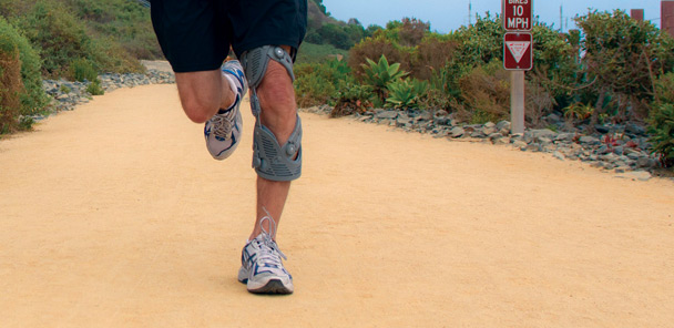
Valgus unloader braces
Despite OA management guidelines recommending interventions that alter the knee mechanical environment,14,15,39-41 new research has found that physicians treat patients with OA by prescribing medications for pain and inflammation or referring for surgical interventions rather than prescribing treatments that address the underlying mechanisms of disease progression.42 Unloader bracing is one conservative management intervention that has potential to alter the mechanical loading environment of the knee joint, yet is underutilized.41,43 Beaudreuil et al41 provide an excellent review of the evidence supporting unloader braces, identifying two areas in need of research. One relates to improving understanding of the biomechanical mechanisms for brace effects, given that inconsistent findings have been presented in the literature; the other determining optimal duration (dose) for brace wear.
Briefly, what we know about biomechanical mechanisms are related primarily to the valgus unloader brace, as it is the type of brace most commonly prescribed for medial compartment knee OA. These braces are designed to apply a three-point force system around the knee joint that results in a net valgus (abduction) knee moment, altering frontal plane alignment and theoretically unloading the medial compartment.44-46 While one study showed minimal effects of a valgus unloader brace on the knee adduction moment,51 the majority of 3D gait analysis studies have shown that valgus unloader braces do reduce the knee adduction moment during stance.44,45,47-50 However, these changes are immediate, and studies of the long-term effects of brace wear are not conclusive.52-54
Biomechanical studies are varied with respect to population and brace type, but improvements in pain47,51,53-60 and function51,52,56,58,59,61 associated with brace wear have consistently been reported in the literature. Unfortunately, little attention has been given to studying mechanical alterations in other planes or muscle characteristics that can impact joint loading. This is important, as it may provide additional information about the mechanism by which the brace works. Different theories are emerging, in particular, those focusing on the role of the neuromuscular system.
In a small sample of individuals with medial compartment knee OA, Ramsey et al found that muscle co-contraction was significantly reduced in early stance phase with the use of neutral alignment and valgus unloader braces (4° of valgus alignment) during walking.46 Based on these results, the researchers hypothesized that the positive effects of braces could be due to increased stabilization, reducing the need for muscle co-contraction. Improved proprioception has also been reported with the use of valgus unloader braces, supporting the theory that bracing has a neuromuscular effect.62
In addition, Matsuno et al54 reported increased quadriceps strength with prolonged brace wear that was accompanied by a decrease in tibiofemoral angle and a concomitant increase in medial joint space. They hypothesized this mechanical change was due to improved knee stability, supporting the mechanism proposed by Ramsey et al.46 Ramsey and Russell63 also evaluated the evidence supporting the use of valgus unloader braces, and while they presented a more positive case for frontal plane alterations than Beaudreuil et al,41 they indicated that more work is needed to understand the effectiveness of braces on the neuromuscular system.
Part of the reason valgus unloader braces are underprescribed in the conservative management of knee OA may relate to ambiguity in prescription instructions. Brace prescription in the research literature has been highly variable, with study participants told to “wear the brace as often as possible,”50,54,59 “wear on a regular basis,”64 “wear for as many hours and for as many days of the week as they wished,”52 “wear during ambulation,”55 “wear for bothersome activities,”56 “wear brace as instructed by physiotherapist or orthopaedic technician,”46,61 and “wear for prolonged standing and sport.”53 In addition to the varied instruction to patients, few studies report the actual duration of brace wear, making it difficult to ascertain the optimal prescription for benefit, and whether there is an amount of brace wear that may be ineffective or even detrimental.
Dose response
One area of concern for clinicians is the belief that long-duration brace use will result in decreased muscle strength. This thought is primarily driven from the literature on functional knee bracing for anterior cruciate ligament injury. Functional knee braces have been associated with thigh muscle atrophy,65 decreased hamstring performance,66 decreased quadriceps torque,67 and premature muscle fatigue.68 Researchers have hypothesized that muscle impairment is a result of the brace straps causing external compression on the muscles, decreasing blood flow and resulting in poor tissue oxygenation.68 Decreases in knee joint muscle strength69 and increased fatigue70 would be detrimental in patients with knee OA, as both have been found to negatively impact knee joint mechanics, in particular the knee adduction moment, during walking.

So the question is whether the length of time the brace is worn per day would have an impact on outcomes, and on muscle strength in particular.
Our recent study71 is one of the first to examine dose response for valgus unloader bracing in which the effects of braces were examined over a wide range of wear durations. The rationale for looking at dose response is that a traditional randomized controlled study (for nonpharmacological studies in which duration of the intervention is prescribed) may be too restrictive for patients, many of whom have comorbidities, who reside in the community where external variables may affect compliance.72 This appears to be the case for brace studies, as poor compliance rates have been noted in the literature.52,61,73,74
To look at the effect of bracing, a more realistic study design may be to have patients record how long they wear the brace and base dose response on the variability of wear times. We used this approach to examine dose response for muscle strength.71
We asked 24 patients with medial compartment knee OA with Kellgren-Lawrence severity scores ranging from 1 to 4 (out of 5) to wear customized valgus unloader braces “as needed” and record daily hours of brace wear and physical activity (step count measured by pedometer) for six months.71 These patients were not on a wait list for total knee arthroplasty and met functional criteria29 (self-reported ability to walk a city block, jog five meters, and climb a flight of 10 stairs in a reciprocal manner).
Average brace wear duration was approximately five hours per day, but there was a wide range (0-14 hours/day). We measured pain, function (subjective and objective), and lower extremity strength at baseline and follow up and determined relationships between changes in these variables and duration of brace wear.
Although not statistically significant, increases of 5% to 13% for four quadriceps and hamstring strength measures were found at six months. Positive relationships were found between duration of brace wear and change in both physical activity and hamstrings muscle strength: the longer the brace was worn, the greater the activity and strength increases. However, the positive relationship between brace-wear dose response and the hamstring strength increase was weak.71
What may be more important is that longer brace wear duration was not associated with decreased strength over the six-month follow-up period.
The lack of decreased muscle strength with high brace use is consistent with the findings of Matsuno et al,54 the only other study looking at the effect of valgus knee bracing on quadriceps muscle strength. This study, however, was in a more advanced, weaker, and older OA group who wore the brace for long daily durations (although actual times were not reported).
Interestingly, our work showed that subjective pain and function improved between baseline and follow up, consistent with the literature,47,51-61 yet there was no dose response for these variables.71 Regardless of how little the brace was worn, perceived pain and function improved, although objective measures of function (walking velocity) did not.
This finding, combined with the positive relationship between brace wear dose and change in physical activity, suggests that there may be different patient-specific strategies for wearing the brace. Our study included younger participants with less severe disease than the study Matsuno et al, but the brace wear use was varied, with the longer wear times reported for the older patients with more severe disease and less active participants. The more active people tended to wear the brace less, perhaps only wearing it for specific activities. Conversely, the less active people tended to wear the brace more and the result was an increase in their physical activity levels.
Only two biomechanics studies have asked participants to record their hours of daily brace wear; these reported mean brace wear durations and standard deviations of 6.9 (4.6)52 and 9 (5)50 hours per day. Hewett et al52 did not find a change in knee adduction moment after six months of brace wear, whereas Draganich et al50 found reduced knee adduction moments after only five weeks. This difference is not likely due to the two-hour difference in brace wear per day, but may be related to the severity of OA in the Hewett study. Hewett’s participants were young, with a mean age of 41 years, and the majority had had a previous injury. Draganich’s study participants were closer in age (50 years) to our study participants, and we included those with primary OA (i.e., not those with OA secondary to an injury).
The findings of these two biomechanical studies, in addition to our study on dose response, suggest that higher brace wear times do not negatively impact muscle strength or knee adduction moment parameters.
Future research
Our single dose response study71 had a relatively brief follow-up period, hence longer term studies are needed. Furthermore, additional work is needed to determine whether dose affects knee joint kinematics and kinetics and muscle activation patterns that have been found to be altered immediately following brace application.44-48,51
We are presently expanding our dose-response analysis to include 3D kinematic and kinetic dynamic waveform data and muscle activation patterns to better understand joint loading throughout the gait cycle. Preliminary findings suggest that changes in specific variables are related to brace wear time. This information will improve our understanding of whether there is an optimal wear time for positive mechanical results.
In addition to looking at dose response of the valgus unloader brace, an important piece of the puzzle is determining which patients are the best candidates for bracing. The variability in response to braces in our study and others implies that there are responders and nonresponders; identifying these individuals and their dose response patterns would be valuable.
Preliminary work from our laboratory has shown that not all individuals respond with a reduced knee adduction moment following brace application. Some individuals experience decreased knee adduction moments during walking with a brace, while others have increased knee adduction moments.75 These differential effects were found in the first two weeks of wearing the valgus unloader brace. Superficially, this might indicate that the brace may actually worsen the mechanical loading environment at the knee in some subgroups. However, the knee adduction moment magnitude does not provide the entire story with respect to the joint loading environment.
A comprehensive 3D motion and force analysis, along with the muscle activation patterns, will provide more complete information. Follow-up studies are needed to determine whether the different responses persist over time and, if so, whether we can identify subgroups of responders and nonresponders with respect to symptoms, joint mechanical characteristics, and structural progression.
Clinical implications
Despite numerous studies showing that valgus unloader braces improve symptoms and evidence that they alter the mechanical environment of the knee, this conservative management option remains underutilized in knee OA management, perhaps because of ambiguity in prescription parameters or concerns about muscle impairment with prolonged use.
While more dose-response studies are needed to determine the optimal duration of brace wear and the effects of bracing on the mechanical loading environment of the knee joint, the available literature indicates that, in terms of pain, function, activity, and muscle strength, there is potential benefit and no harm in wearing the brace as needed, even for prolonged periods of time.
Cheryl Hubley-Kozey, PhD, is a professor in the schools of Physiotherapy and Biomedical Engineering at Dalhousie University in Halifax, Canada, whose research focus is on improving function of those with musculoskeletal conditions. Gillian Hatfield Murdock, PT, MSc, is a physiotherapist and doctoral candidate in the School of Biomedical Engineering at Dalhousie who is interested in the relationship between physical activity and knee OA progression.
Acknowledgements: Canadian Institutes of Health and Nova Scotia Health Research Foundation.
1. Bombardier C, Hawker G, Mosher D, et al. Arthritis Alliance of Canada. The impact of arthritis in Canada: Today and over the next 30 years. http://www.arthritisalliance.ca/docs/20111022_2200_impact_of_arthritis.pdf. Published October 27, 2011. Accessed July 31, 2012.
2. Lane NE, Brandt K, Hawker G, et al. OARSI-FDA initiative: Defining the disease state of osteoarthritis. Osteoarthritis Cartilage 2011;19(5):478-482.
3. Lawrence RC, Felson DT, Helmick CG, et al. Estimates of the prevalence of arthritis and other rheumatic conditions in the United States. Part II. Arthritis Rheum 2008;58(1):26-35.
4. Brandt KD, Radin EL, Dieppe PA, van de Putte L. Yet more evidence that osteoarthritis is not a cartilage disease. Ann Rheum Dis 2006;65(10):1261-1264.
5. Yelin E, Murphy L, Cisternas MG, et al. Medical care expenditures and earnings losses among persons with arthritis and other rheumatic conditions in 2003, and comparisons with 1997. Arthritis Rheum 2007;56(5):1397-1407.
6. Hunter DJ, Felson DT. Osteoarthritis. BMJ 2006;332(7542):639-642.
7. Canadian Joint Replacement Registry. 2006 report: Hip and knee replacements in Canada. Canadian Institute for Health Information website. https://secure.cihi.ca/free_products/CJRR_Annual_Report_Hip_Knee_Replacements_2006_e.pdf. Published 2006. Accessed July 31, 2012.
8. NIH consensus statement on total knee replacement. NIH Consens State Sci Statements 2003;20(1):1-34.
9. Buckwalter JA, Stanish WD, Rosier RN, et al. The increasing need for nonoperative treatment of patients with osteoarthritis. Clin Orthop Relat Res 2001;(385):36-45.
10. Ding C, Cicuttini F, Jones G. Do NSAIDs affect longitudinal changes in knee cartilage volume and knee cartilage defects in older adults? Am J Med 2009;122(9):836-842.
11. Reijman M, Bierma-Zeinstra SM, Pols HA, et al. Is there an association between the use of different types of nonsteroidal antiinflammatory drugs and radiologic progression of osteoarthritis? The Rotterdam study. Arthritis Rheum 2005;52(10):3137-3142.
12. Henriksen M, Simonsen EB, Alkjaer T, et al. Increased joint loads during walking—a consequence of pain relief in knee osteoarthritis. Knee 2006;13(6):445-450.
13. Hurwitz DE, Ryals AR, Block JA, et al. Knee pain and joint loading in subjects with osteoarthritis of the knee. J Orthop Res 2000;18(4):572-579.
14. Zhang W, Nuki G, Moskowitz RW, et al. OARSI recommendations for the management of hip and knee osteoarthritis, part II: OARSI evidence-based, expert consensus guidelines. Osteoarthritis Cartilage 2008;16(2):137-162.
15. Zhang W, Nuki G, Moskowitz RW, et al. OARSI recommendations for the management of hip and knee osteoarthritis: Part III: Changes in evidence following systematic cumulative update of research published through January 2009. Osteoarthritis Cartilage 2010;18(4):476-499.
16. Brandt KD, Mazzuca SA. Experience with a placebo-controlled randomized clinical trial of a disease-modifying drug for osteoarthritis: The doxycycline trial. Rheum Dis Clin North Am 2006;32(1):217-234.
17. Hoemann CD, Sun J, McKee MD, et al. Chitosan-glycerol phosphate/blood implants elicit hyaline cartilage repair integrated with porous subchondral bone in microdrilled rabbit defects. Osteoarthritis Cartilage 2007;15(1):78-89.
18. Radin EL, Martin RB, Burr DB, et al. Effects of mechanical loading on the tissues of the rabbit knee. J Orthop Res 1984;2(3):221-34.
19. Walker ER, Boyd RD, Wu DD, et al. Morphologic and morphometric changes in synovial membrane associated with mechanically induced osteoarthrosis. Arthritis Rheum 1991;34(5):515-524.
20. O’Connor BL, Brandt KD. Neurogenic factors in the etiopathogenesis of osteoarthritis. Rheum Dis Clin North Am 1993;19(3):581-605.
21. Andriacchi TP, Mündermann A, Smith RL, et al. A framework for the in vivo pathomechanics of osteoarthritis at the knee. Ann Biomed Eng 2004;32(3):447-457.
22. Andriacchi TP, Mündermann A. The role of ambulatory mechanics in the initiation and progression of knee osteoarthritis. Curr Opin Rheumatol 2006;18(5):514-518.
23. Miyazaki T, Wada M, Kawahara H, et al. Dynamic load at baseline can predict radiographic disease progression in medial compartment knee osteoarthritis. Ann Rheum Dis 2002;61(7):617-622.
24. Hubley-Kozey C, Deluzio K, Dunbar M. Muscle co-activation patterns during walking in those with severe knee osteoarthritis. Clin Biomech 2008;23(1):71-80.
25. Astephen JL, Deluzio KJ, Caldwell GE, et al. Gait and neuromuscular pattern changes are associated with differences in knee osteoarthritis severity levels. J Biomech 2008;41(4):868-876.
26. Zeni JA Jr, Higginson JS. Differences in gait parameters between healthy subjects and persons with moderate and severe knee osteoarthritis: A result of altered walking speed? Clin Biomech 2009;24(4):372-378.
27. Landry SC, McKean KA, Hubley-Kozey CL, et al. Knee biomechanics of moderate OA patients measured during gait at a self-selected and fast walking speed. J Biomech 2007;40(8):1754-1761.
28. Hubley-Kozey CL, Deluzio KJ, Landry SC, et al. Neuromuscular alterations during walking in persons with moderate knee osteoarthritis. J Electromyogr Kinesiol 2006;16(4):365-378.
29. Hubley-Kozey CL, Hill NA, Rutherford DJ, et al. Co-activation differences in lower limb muscles between asymptomatic controls and those with varying degrees of knee osteoarthritis during walking. Clin Biomech 2009;24(5):407-414.
30. Thorp LE, Sumner DR, Block JA, et al. Knee joint loading differs in individuals with mild compared with moderate medial knee osteoarthritis. Arthritis Rheum 2006;54(12):3842-3849.
31. Ahlbäck S. Osteoarthrosis of the knee. A radiographic investigation. Acta Radiol Diagn 1968;Suppl 277:7-72.
32. Frontera WR, Silver JK. Essentials of physical medicine and rehabilitation. Philadelphia: Hanley & Belfus; 2002.
33. Astephen JL, Deluzio KJ, Caldwell GE, Dunbar MJ. Biomechanical changes at the hip, knee, and ankle joints during gait are associated with knee osteoarthritis severity. J Orthop Res 2008;26(3):332-341.
34. Mündermann A, Dyrby CO, Andriacchi TP. Secondary gait changes in patients with medial compartment knee osteoarthritis: Increased load at the ankle, knee, and hip during walking. Arthritis Rheum 2005;52(9):2835-2844.
35. Gross KD, Hillstrom HJ. Noninvasive devices targeting the mechanics of osteoarthritis. Rheum Dis Clin North Am 2008;34(3):755-776.
36. Rutherford DJ, Hubley-Kozey CL, Stanish WD, Dunbar MJ. Neuromuscular alterations exist with knee osteoarthritis presence and severity despite walking velocity similarities. Clin Biomech 2011;26(4):377-383.
37. Astephen Wilson JL, Deluzio KJ, Dunbar MJ, et al. The association between knee joint biomechanics and neuromuscular control and moderate knee osteoarthritis radiographic and pain severity. Osteoarthritis Cartilage 2011;19(2):186-193.
38. Zeni JA, Rudolph K, Higginson JS. Alterations in quadriceps and hamstrings coordination in persons with medial compartment knee osteoarthritis. J Electromyogr Kinesiol 2010;20(1):148-154.
39. Felson DT. Clinical practice. Osteoarthritis of the knee. N Engl J Med 2006;354(8):841-848.
40. Recommendations for the medical management of osteoarthritis of the hip and knee: 2000 update. American College of Rheumatology Subcommittee on Osteoarthritis Guidelines. Arthritis Rheum 2000;43(9):1905-1915.
41. Beaudreuil J, Bendaya S, Faucher M, et al. Clinical practice guidelines for rest orthosis, knee sleeves, and unloading knee braces in knee osteoarthritis. Joint Bone Spine 2009;76(6):629-636.
42. Hunter DJ. Osteoarthritis. Best Pract Res Clin Rheumatol 2011;25(6):801-814.
43. Hand L. OA knee bracing in family practice. LER: Resource Guide 2011;3(12):70.
44. Pollo FE, Otis JC, Backus SI, et al. Reduction of medial compartment loads with valgus bracing of the osteoarthritic knee. Am J Sports Med 2002;30(3):414-421.
45. Fantini Pagani CH, Potthast W, Brüggemann GP. The effect of valgus bracing on the knee adduction moment during gait and running in male subjects with varus alignment. Clin Biomech 2010;25(1):70-76.
46. Ramsey DK, Briem K, Axe MJ, Snyder-Mackler L. A mechanical theory for the effectiveness of bracing for medial compartment osteoarthritis of the knee. J Bone Joint Surg Am 2007;89(11):2398-2407.
47. Lindenfeld TN, Hewett TE, Andriacchi TP. Joint loading with valgus bracing in patients with varus gonarthrosis. Clin Orthop Relat Res 1997;(344):290-297.
48. Self BP, Greenwald RM, Pflaster DS. A biomechanical analysis of a medial unloading brace for osteoarthritis in the knee. Arthritis Care Res 2000;13(4):191-197.
49. Toriyama M, Deie M, Shimada N, et al. Effects of unloading bracing on knee and hip joints for patients with medial compartment knee osteoarthritis. Clin Biomech 2011;26(5):497-503.
50. Draganich L, Reider B, Rimington T, et al. The effectiveness of self-adjustable custom and off-the-shelf bracing in the treatment of varus gonarthrosis. J Bone Joint Surg Am 2006;88(12):2645-2652.
51. Gaasbeek RD, Groen BE, Hampsink B, et al. Valgus bracing in patients with medial compartment osteoarthritis of the knee. A gait analysis study of a new brace. Gait Posture 2007;26(1):3-10.
52. Hewett TE, Noyes FR, Barber-Westin SD, Heckmann TP. Decrease in knee joint pain and increase in function in patients with medial compartment arthrosis: A prospective analysis of valgus bracing. Orthopedics 1998;21(2):131-138.
53. Horlick SG, Loomer RL. Valgus knee bracing for medial gonarthrosis. Clin J Sports Med 1993;3(4):251-255.
54. Matsuno H, Kadowaki KM, Tsuji H. Generation II knee bracing for severe medial compartment osteoarthritis of the knee. Arch Phys Med Rehabil 1997;78(7):745-749.
55. Katsuragawa Y, Fukui N, Nakamura K. Change of bone mineral density with valgus knee bracing. Int Orthop 1999;23(3):164-167.
56. Kirkley A, Webster-Bogaert S, Litchfield R, et al. The effect of bracing on varus gonarthrosis. J Bone Joint Surg Am 1999;81(4):539-548.
57. Komistek RD, Dennis DA, Northcut EJ, et al. An in vivo analysis of the effectiveness of the osteoarthritic knee brace during heel-strike of gait. J Arthroplasty 1999;14(6):738-742.
58. Barnes CL, Cawley PW, Hederman B. Effect of CounterForce brace on symptomatic relief in a group of patients with symptomatic unicompartmental osteoarthritis: A prospective 2-year investigation. Am J Orthop 2002;31(7):396-401.
59. Richards JD, Sanchez-Ballester J, Jones RK, et al. A comparison of knee braces during walking for the treatment of osteoarthritis of the medial compartment of the knee. J Bone Joint Surg Br 2005;87(7):937-939.
60. Dennis DA, Komistek RD, Nadaud MC, Mahfouz M. Evaluation of off-loading braces for treatment of unicompartmental knee arthrosis. J Arthroplasty 2006;21(4 Suppl 1):2-8.
61. Brouwer RW, van Raaij TM, Verhaar JA, et al. Brace treatment for osteoarthritis of the knee: A prospective randomized multi-centre trial. Osteoarthritis Cartilage 2006;14(8):777-783.
62. Birmingham TB, Kramer JF, Kirkley A, et al. Knee bracing for medial compartment osteoarthritis: Effects on proprioception and postural control. Rheumatology 2001;40(3):285-289.
63. Ramsey DK, Russell ME. Unloader braces for medial compartment knee osteoarthritis: Implications on mediating progression. Sports Health 2009;1(5):416-426.
64. Davidson PL, Sanderson DJ, Loomer RL. Kinematics of valgus bracing for medial gonarthrosis: Technical report. Clin Biomech 1998;13(6):414-419.
65. Risberg MA, Holm I, Steen H, et al. The effect of knee bracing after anterior cruciate ligament reconstruction. A prospective, randomized study with two years’ follow-up. Am J Sports Med 1999;27(1):76-83.
66. Wojtys EM, Kothari SU, Huston LJ. Anterior cruciate ligament functional brace use in sports. Am J Sports Med 1996;24(4):539-546.
67. Houston ME, Goemans PH. Leg muscle performance of athletes with and without knee support braces. Arch Phys Med Rehabil 1982;63(9):431-432.
68. Styf J. The effects of functional knee bracing on muscle function and performance. Sports Med 1999;28(2):77-81.
69. Sturnieks DL, Besier TF, Hamer PW, et al. Knee strength and knee adduction moments following arthroscopic partial meniscectomy. Med Sci Sports Exerc 2008;40(6):991-997.
70. Murdock GH, Hubley-Kozey CL. Effect of a high intensity quadriceps fatigue protocol on knee joint mechanics and muscle activation during gait in young adults. Eur J Appl Physiol 2012;112(2):439-449.
71. Hurley ST, Hatfield Murdock GL, Stanish WD, Hubley-Kozey CL. Is there a dose response for valgus unloader brace usage on knee pain, function, and muscle strength? Arch Phys Med Rehabil 2012;93(3):496-502.
72. Hurley MV. Muscle dysfunction and effective rehabilitation of knee osteoarthritis: What we know and what we need to find out. Arthritis Rheum 2003;49(3):444-452.
73. Giori NJ. Load-shifting brace treatment for osteoarthritis of the knee: A minimum 2 1/2-year follow-up study. J Rehabil Res Dev 2004;41(2):187-194.
74. Wilson B, Rankin H, Barnes CL. Long-term results of an unloader brace in patients with unicompartmental knee osteoarthritis. Orthopedics 2011;34(8):e334-e337.
75. Conrad J, Hubley-Kozey CL, Astephen-Wilson JL, et al. The correction of medial joint loading with valgus unloader brace is related to varus thrust. In: Proceedings from the Canadian Orthopaedic Research Society; June 2012; Ottawa, Canada. Abstract 93.


I disagree with the assumptions that long term use of an unloader brace will not impact muscle strength. I am now undergoing PT to try to regain some of the loss muscle strength – as I can no longer climb stairs without using the rails to pull myself up. I have lost a lot in my quads in that leg.
That is the negative. Here is the positive. I wear the brace when I am going to be physically active -eg exercise classes, swimming (5x a week), going shopping etc. Average use is not going to exceed 3 or 4 hours a day – some days I don’t wear it at all.
I also have severe osteoarthritis – right lateral and medial compartment. My GP suggested an Ossur brace 2006 – My otho said okay but I still needed surgery someday. I am now 74. My brace is now 12 and I love it. Mostly pain free – 5 tylenol a week max. No plans for surgery ever. But I really want to regain a lot of the muscle strength – quads – that I have lost throughout the years.