
 By Howard Kashefsky, DPM, FACFAS
Sponsored by an educational grant from Medi USA.
Metatarsalgia is a common foot disorder. The term metatarsalgia refers to a pain syndrome in the forefoot and not to a specific diagnosis.1 Pain is confined to the area across the plantar forefoot, including the second through fourth metatarsal heads. However, it is frequently accompanied by deformity of the first and fifth rays, as well as the toes.2
Many different diagnoses have been identified as the cause of the painful forefoot. Excessive stress may result in ligamentous strain, synovitis, capsulitis, stress fracture, or degenerative arthritis. Metatarsalgia may be associated with a tight Achilles’ tendon (equinus), cerebral palsy, stroke, multiple sclerosis, or other neurological diseases. Metatarsalgia also results from pathological alterations in forefoot structure due to hallux valgus, hallux limitus, rheumatoid arthritis, osteomyelitis, or osteochondrosis (Freiberg disease). Circulatory or metabolic disorders may also be associated with metatarsalgia.3-8 Brachymetatarsia, an arrest of normal metatarsal growth and development with a resultant short ray, may cause symptoms.9 A Morton neuroma or other nerve injuries may be associated with forefoot pain.10 Hammertoes can occur in association with joint dislocation, and may contribute.11
Cavus foot and pes planus foot types have both been associated with metatarsalgia. Cavus foot type as well as pes planus with hypermobile first and fifth rays have been associated with increased shearing at the forefoot.12,13 Metatarsal weightbearing is increased with the cavus foot because a disproportionate amount of weight is borne by the heel and forefoot. It is not uncommon to observe retracted or clawed toes with cavus feet, which decreases the ability to unload the metatarsals at push-off.14 The pes planus foot type (resulting from genu valgum, rearfoot valgus, or forefoot varus) remains pronated during midstance and inhibits proper supination, which compromises the propulsive function of the forefoot.
Although plantar fat pad loss makes sense intuitively as a cause of metatarsalgia, a study by Waldecker suggests the two may not be associated; more research needs to be done.15 Unintended iatrogenic metatarsalgia after bunion surgery can be the result of a short first metatarsal or elevation of the first ray.16,17
Classifications
Helal et al classified metatarsalgia as either primary or secondary.18 Primary metatarsalgia is structural—an anatomical abnormality resulting in increased pressure under the metatarsal heads. Examples include hallux rigidus, long or short metatarsal bones, and possibly pes cavus. Treatment should be focused on offloading the metatarsal heads and should mechanically direct force away from the point of pressure.
Secondary metatarsalgia is defined as pain that does not originate within the metatarsal area. Conditions such as rheumatoid arthritis, sesamoiditis, and equinus can all lead to localized pain at the ball of the foot. The origin of metatarsalgia can be multifactorial. Scranton found that 31 of 98 patients had two or more mechanical etiologies for primary metatarsalgia, and that often, primary and secondary metatarsalgia existed together.19 Effective treatment will address the area of pain, the function of the foot, and, if necessary, the systemic disease.
Scranton found 23 different diagnoses of metatarsalgia in 98 patients. Forty-five patients had primary metatarsalgia, 12 of whom had static disorders and 12 of whom had iatrogenic (postoperative) etiologies. Thirty-three patients had secondary metatarsalgia, 11 of whom had rheumatoid arthritis and 10 of whom had sesamoiditis. Twenty patients experienced pain under the forefoot.19
Viladot has classified metatarsal pathomechanics as an overload of anterior support or an irregular distribution of the metatarsal load.20 Irregular metatarsal load syndromes are further separated into four groups: first ray overload, first ray insufficiency, central ray overload, and central ray insufficiency.
 Metatarsalgia in athletes
Metatarsalgia is common in sports, including rock climbing, running, and cycling. A potential cause of these injuries is excessive plantar pressure in the forefoot region.21,22
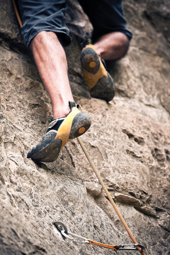
In particular, pain associated with metatarsal stress fracture has plagued military personnel throughout history but has now become more common in the civilian population with the increasing popularity of recreational long-distance running, aerobics, and jumping sports.23,24 Metatarsal stress fractures and metatarsalgia also are fairly common among competitive athletes, especially runners.25-27 Buda et al identified metatarsalgia in 12.5% of 144 rock climbers.28
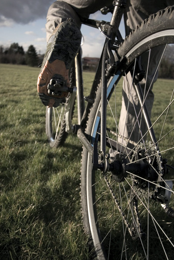
Cycling is associated with metatarsalgia.29 Carbon fiber cycling shoes have become popular for their stiffness, but the stiffer material is also associated with 18% higher peak plantar pressures in the forefoot, leading Jarboe and Quesada to recommend that competitive or professional cyclists suffering from metatarsalgia or ischemia should be especially careful not to aggravate these foot conditions.30
Treatment
Multiple studies support the use of treatment focused on reducing forefoot plantar pressure.31-34 Postema et al reported a reduction in forefoot plantar pressure and force impulse both with a rocker sole and with custom foot orthoses in individuals with a history of metatarsalgia.32 Individuals with current pain preferred a custom-molded insole more often than those without pain.
The efficacy of rocker shoes for reducing forefoot plantar pressure during walking has been well documented in both healthy individuals and patients with forefoot problems such as metatarsalgia.35-39 Sobhani et al found that running in rocker shoes was associated with a significant reduction in all pressure parameters in the central and lateral forefoot as well as reduced force time integral and maximum mean pressure in the medial forefoot.40 Although the study participants (all healthy runners) found the rocker shoes less comfortable than standard running shoes, the authors concluded rocker shoes might be beneficial for runners who are recovering from metatarsalgia or stress fractures in the forefoot region.
Treatment with metatarsal pads is a common standalone therapy for metatarsalgia. A 1990 study by Holmes and Timmerman used pedobarography to determine the effect of metatarsal pads on pressure under the metatarsal heads in 10 asymptomatic volunteers.41 They found that the met pads were associated with significantly reduced pressure at the second metatarsal head for all participants; pressure reduction with met pad use decreased for each subsequent lesser metatarsal head lateral to the second metatarsal.
Another study of metatarsal pads, published in 1994, expanded on this idea by measuring the intervention’s effect on peak pressures in eight discrete plantar locations on the hindfoot, midfoot, and forefoot.42 In 10 asymptomatic individuals, met pad use was associated with statistically significant increases in plantar pressure at the metatarsal shaft region, suggesting offloading of the metatarsal heads. Although there were no statistically significant changes in any other plantar region, there was a mild decrease in pressure at the first and second metatarsal heads and slight increases laterally. In addition, contact duration decreased at all metatarsal head locations, and pressure-time integral (PTI) decreased at the first, second, third, and fourth metatarsals.
Location of met pads seems to be crucial for plantar pressure reduction, as a research group in Taiwan demonstrated.43 Metatarsal pads, each measuring 55 mm in length, 36 mm in width, and 10 mm in height, were uniformly used in 10 individuals with a history of primary metatarsalgia. The greatest pressure reduction was achieved when the metatarsal pad was placed just proximal to the site of peak metatarsal head pressure. The study revealed positional differences as small as 4 mm could influence the metatarsal pad’s ability to reduce plantar pressures at the metatarsal heads. However, for clinicians to use the same type of assessment to place a metatarsal pad for each patient is impractical.

Because one of the most important outcomes of treatment is pain relief, in 2006 the same Taiwanese research group assessed the correlation between the use of a metatarsal pad and subjective symptoms.44 Thirteen patients with secondary metatarsalgia wore metatarsal pads positioned under and just proximal to the second metatarsal head for two weeks. Improvements in visual analog pain scores were statistically correlated with reduction in PTI and, more strongly, with reduction in maximum peak pressure.
 Additional studies of secondary metatarsalgia have focused primarily on pain relief with the use of met pads in combination with foot orthoses in patients with rheumatoid arthritis.45,46 These studies found that metatarsal pads were associated with statistically significant decreases in peak plantar pressures, pressure-time intervals, and patients’ pain, along with increased quality of life. These studies also found the use of a custom orthosis without the pad provided a frequent decrease of peak pressures and decreased metatarsal pain in varying amounts.
In a 2006 study of 20 patients with diabetes, Mueller et al found that both a total contact insert and a metatarsal pad were associated with statistically significant reduction of pressure under the metatarsal heads.47 Patients were analyzed while wearing just a shoe, a shoe with total contact orthoses, and a shoe with the same orthoses but with a metatarsal pad added. The total contact orthoses in this study increased the foot contact area by an average of 27%, primarily in the arch. The orthoses reduced metatarsal peak pressures by 19% to 24% and PTI by 16% to 23%. The addition of a metatarsal pad, although it did not increase contact area, reduced peak pressure by an additional 15% to 20%, and PTI by an additional 22% to 32%. The authors reported that adding the metatarsal pad increased the pressure peak at the second metatarsal shaft by an amazing 308%, indicating that, although the pad did not increase the surface area, it redistributed pressure from the metatarsal head to the shaft.
Treatment of metatarsalgia with foot orthoses alone has also been researched. The purpose of orthotic therapy is to increase the patient’s tolerance of weightbearing, facilitate proper foot function, and normalize gait. The orthosis should balance weight distribution across the metatarsals by correcting or compensating for any biomechanical malalignments that alter foot biomechanics.
Orthotic design considerations
Orthotic design should be tailored to each individual case. The disproportionately high weightbearing areas in the cavus foot can be reduced by adding support along the lateral and medial longitudinal arches.14 The pes planus foot requires control of forefoot or rearfoot varus by medial posting.48 Medial column instability due to excessive rearfoot pronatory motion often leads to dorsiflexion of the first ray and subsequent overloading of the second, limiting the first metatarsophalangeal joint motion.49 A wide orthotic plate made from a cast with the first ray plantar flexed helps maintain contact with the more medial aspect of the foot, allows greater motion of the big toe, and enhances weightbearing under the first metatarsal head, decreasing pressure under the second metatarsal head.49
In general, the following steps14 established in 1985 are still valid for orthotic management of metatarsalgia:
- Perform a complete clinical examination;
- Establish a diagnosis;
- Determine which orthotic features will help achieve treatment goals;
- Fabricate the device from the correct materials;
- Determine the therapeutic benefit of each of the device’s features;
- Analyze the effect of the device on the patient’s gait; and
- Reevaluate the orthosis periodically.
Custom versus ready-made orthoses for metatarsalgia have been studied. Kelly and Winson found both custom and prefabricated insoles were associated with reduced plantar forefoot pressure in patients with lesser metatarsalgia, however, the custom group reported a 16% higher compliance rate and greater self-reported symptom improvement.50
Ki et al found orthoses with minimum arch fill were far more effective than flat insoles for redistributing peak plantar pressures in 30 healthy volunteers.51 They also demonstrated that, the greater the contact in the arch area, the greater the decrease in rearfoot and forefoot pressures. The paper also revealed that custom devices made using either CAD-CAM technology or foam impression were far superior to flat insoles for reducing forefoot plantar pressures.
Orthotic therapy should be used only with suitable footwear. Metatarsalgia patients should use shoes with a low heel, stiff sole, and large, rounded toe box.14,52-54
A variety of orthotic approaches may be used to alter the weightbearing load at the metatarsals. Shaft padding relieves the symptomatic metatarsal head by transferring the load to the proximal metatarsal shaft. The first, second, and fifth metatarsals can be padded with flexible materials, while the third and fourth metatarsals can be supported with pear-shaped metatarsal pads.55-57 The success of shaft padding requires placing the support just proximal to the metatarsal head and not underneath it.
A topcover and a forefoot extension can be used to add cushioning, which dampens force. Research suggests softer topcover materials give patients more comfort and increase their tolerance of orthotic devices,58 which may have implications for multiple pathologies. Materials such as closed-cell neoprene, plastazote, and Poron can add shock absorption under the forefoot.
Topcovers require periodic replacement, dependent on patient weight, moisture, and frequency of use. Since compression of very soft materials over time negates the benefits of shock absorption the patient needs, noncompliance and loss of effectiveness can result if these materials are not replaced. Poron extensions that maintain their ability to cushion the metatarsal heads also attenuate pressure by delaying compression.
Howard Kashefsky, DPM, is the director of podiatry services at the University of North Carolina Hospitals in Chapel Hill.
- Shoji H. The foot and ankle. In: D’Ambrosia DD, ed. Musculoskeletal disorders regional examination and differential diagnosis. Philadelphia: JB Lippincott Co.;1977: 509-512.
- Espinosa N, Brodsky JW, Maceira E. Metatarsalgia. J Am Acad Orthop Surg 2010;18(8):474-485.
- Turek SL. Orthopaedics: Principles and their applications. Philadelphia: JB Lippincott Co; 1977.
- Dinoá V, von Ranke F, Costa F, Marchiori E. Evaluation of lesser metatarsophalangeal joint plantar plate tears with contrast-enhanced and fat-suppressed MRI. Skeletal Radiol 2016;45(5):635-644.
- Slullitel G, López V, Calvi JP, et al. Effect of first ray insufficiency and metatarsal index on metatarsalgia in hallux valgus. Foot Ankle Int 2016;37(3):300-306.
- Cychosz CC, Phisitkul P, Belatti DA, et al. Gastrocnemius recession for foot and ankle conditions in adults: Evidence-based recommendations. Foot Ankle Surg 2015;21(2):77-85.
- Cazeau C, Stiglitz Y. Effects of gastrocnemius tightness on forefoot during gait. Foot Ankle Clin 2014;19(4):649-657.
- Maddali Bongi S, Del Rosso A, Mikhaylova S, et al. A comparison of two podiatric protocols for metatarsalgia in patients with rheumatoid arthritis and osteoarthritis. Clin Exp Rheumatol 2014;32(6):855-863.
- Pandey PK, Pawar I, Beniwal SK, Verma RR. Brachymetatarsia with accessory navicular in right foot: A rare coincidental finding. Chin J Traumatol 2016;19(1):56-58.
- Bauer T, Gaumetou E, Klouche S, et al. Metatarsalgia and Morton’s disease: comparison of outcomes between open procedure and neurectomy versus percutaneous metatarsal osteotomies and ligament release with a minimum of 2 years of follow-up. J Foot Ankle Surg 2015;54(3):373-377.
- DiPreta JA. Metatarsalgia, lesser toe deformities, and associated disorders of the forefoot. Med Clin North Am 2014;98(2):233-251.
- Cailliet R. Soft tissue pain and disability. Philadelphia: FA Davis; 1977.
- Neale D, Hooper G. Clowes C, Whiting MF. Adult foot disorders. In: Neale D, ed. Common foot disorders: Diagnosis and management. Edinburgh: Churchill Livingstone; 1981: 69-70.
- Doxey G. Management of metatarsalgia with foot orthotics. J Orthop Sports Phys Ther 1985;6(6):324-333.
- Waldecker U. Plantar fat pad atrophy: a cause of metatarsalgia? J Foot Ankle Surg 2001;40(1):21-27.
- Barouk P. Recurrent metatarsalgia. Foot Ankle Clin 2014;19(3):407-424.
- Maceira E, Monteagudo M. Transfer metatarsalgia post hallux valgus surgery. Foot Ankle Clin 2014;19(2):285-307.
- Helal B, Thomas N, Nissen KI. Disorders of the lesser ray. In: Helal B, Wilson D, eds. The foot. Edinburugh: Churchill Livingstone: 1988:486.
- Scranton PE Jr. Metatarsalgia: diagnosis and treatment. J Bone Joint Surg 1980;62(5):723-732.
- Viladot A. The metatarsals. In: Jahss MH, ed. Disorders of the foot, Vol 1. Philadelphia: WB Saunders; 1982:659-710.
- Bardelli M, Turelli L, Scoccianti G. Definition and classification of metatarsalgia. Foot Ankle Surg 2003;9(2):79-85.
- Nagel A, Fernholz F, Kibele C, Rosenbaum D. Long distance running increases plantar pressures beneath the metatarsal heads: a barefoot walking investigation of 200 marathon runners. Gait Posture 2008;27(1):152-155.
- McBryde AM Jr. Stress fractures in athletes. J Sports Med 1975;3(5):212-217.
- Brown TD, Micheli LJ. Foot and ankle injuries in dance. Am J Orthop 2004;33(6):303-309.
- Sobhani S, Dekker R, Postema K, Dijkstra PU. Epidemiology of ankle and foot overuse injuries in sports: a systematic review. Scand J Med Sci Sports 2103;23(6):669-686.
- Glasoe WM, Allen MK, Kepros T, et al. Dorsal first ray mobility in women athletes with a history of stress fracture of the second or third metatarsal. J Orthop Sports Phys Ther 2002;32:560-565.
- Bennell KL, Malcolm SA, Thomas SA, et al. The incidence and distribution of stress fractures in competitive track and field athletes. A twelve-month prospective study. Am J Sports Med 1996;24(2):211-217.
- Buda R, Di Caprio F, Bedetti L, et al. Foot overuse diseases in rock climbing: an epidemiologic study. J Am Podiatr Med Assoc 2013;103(2):113-120.
- Wanich T, Hodgkins C, Columbier JA, et al. Cycling injuries of the lower extremity. J Am Acad Orthop Surg 2007;15(12):748-756.
- Jarboe NE, Quesada PM. The effects of cycling shoe stiffness on forefoot pressure. Foot Ankle Int 2003;24(10):784-788.
- Hassouna H, Singh D. Morton’s metatarsalgia: pathogenesis, aetiology and current management Acta Orthop Belg 2005;71(6):646-655.
- Postema K, Burm PE, Zande ME, Limbeek JV. Primary metatarsalgia: the influence of a custom moulded insole and a rockerbar on plantar pressure. Prosthet Orthot Int 1998;22(1):35-44.
- Espinosa E, Maceira M, Myerson MS. Current concept review: metatarsalgia. Foot Ankle Int 2008;29(8):871-879.
- Deshaies A, Roy P, Symeonidis P, et al. Metatarsal bars more effective than metatarsal pads in reducing impulse on the second metatarsal head. Foot 2011;21(4):172-175.
- Schaff PS, Cavanagh PR. Shoes for the insensitive foot: the effect of a rocker bottom shoe modification on plantar pressure distribution. Foot Ankle 1990;11(3):129-140.
- Brown D, Wertsch JJ, Harris GF, et al. Effect of rocker soles on plantar pressures Arch Phys Med Rehabil 2004;85(1):81-86.
- Fuller E, Schroeder S, Edwards J. Reduction of peak pressure on the forefoot with a rigid rocker-bottom postoperative shoe. J Am Podiatr Med Assoc 2001;91(10):501-507.
- Kavros S, Van Straaten M, Coleman Wood K, Kaufman K. Forefoot plantar pressure reduction of off-the-shelf rocker bottom provisional footwear. Clin Biomech 2011;26(7):778-782.
- Praet SF, Louwerens JW. The influence of shoe design on plantar pressures in neuropathic feet. Diabetes Care 2003;26(2):441-445.
- Sobhani S, van den Heuvel E, Bredeweg S, et al. Effect of rocker shoes on plantar pressure pattern in healthy female runners. Gait Posture 2014;39(3):920-925.
- Holmes GB Jr, Timmerman L. A quantitative assessment of the effect of metatarsal pads on plantar pressures. Foot Ankle Int 1990;11(3):141-145.
- Chang AH, Abu-Faraj ZU, Harris GF, et al. Multistep measurement of plantar pressure alterations using metatarsal pads. Foot Ankle Int 1994;15(12):654-660.
- Hsi WL, Kang JH, Lee XX. Optimum position of metatarsal pad in metatarsalgia for pressure relief. Am J Phys Med Rehabil 2005;84(7):514-520.
- Kang JH, Chen MD, Chen SC, Hsi WL. Correlations between subjective treatment responses and plantar pressure parameters of metatarsal pad treatment in metatarsalgia patients: a prospective study. BMC Musculoskelet Disord 2006;7:95.
- Hodge MC, Bach TM, Carter GM. Orthotic management of plantar pressure and pain in rheumatoid arthritis. Clin Biomech 1999;14(8):567-575.
- de P Magalhaes E, Davitt M, Filho DJ, et al. The effect of foot orthoses in rheumatoid arthritis. Rheumatology 2006;45(4):449-453.
- Mueller MJ, Lott DJ, Hastings MK, et al. Efficacy and mechanism of orthotic devices to unload metatarsal heads in people with diabetes and a history of plantar ulcers. Phys Ther 2006;86(6):833-842.
- Cracchiolo A 3rd. Management of the arthritic forefoot. Foot Ankle 1982;3(1):17-23.
- Scherer PR, Sanders J, Eldredge, DE, et al. Effect of functional foot orthoses on first metatarsophalangeal joint dorsiflexion in stance and gait. J Am Podiatr Med Assoc 2006;96(6):474-481.
- Kelly A, Winson I. Use of ready-made insoles in the treatment of lesser metatarsalgia: a prospective randomized controlled trial. Foot Ankle Int 1998;19(4):217-220
- Ki SW, Leung AK, Li AN. Comparison of plantar pressure distribution patterns between foot orthoses provided by the CAD-CAM and foam impression methods. Prosthet Orthot Int 2008;32(3):356-362.
- Glass MK, Karno ML, Sella EJ, Zelezmik R. An office based system in the treatment of the arthritic foot. Foot Ankle 1982;3(1):37-40.
- Grundy M, Tosh PA. McLeish RD, Schmidt L. An investigation of the centres of pressure under the foot while walking. J Bone Joint Surg (Br) 1975;57(1):98-103.
- Scranton PE, Pedegana LR, White JP. Gait analysis alterations in support phase forces using supportive devices. Am J Sports Med 1982;10(1):6-11.
- Milgram JE. Padding and devices to relieve the painful foot. In: Jahss MH, ed. Disorders of the foot, Vol 2. Philadelphia: WB Saunders; 1982:1705-1708.
- Cracchiolo A 3rd. Office practice footwear and orthotic therapy. Foot Ankle 1982;2(4):242-248.
- Mueller MJ, Lott DJ, Hastings MK, et al. Efficacy and mechanism of orthotic devices to unload metatarsal heads in people with diabetes and a history of plantar ulcers. Phys Ther 2006;86(6):833-842.
- Pawelka S, Kopf A, Zwick E, et al. Comparison of two insole materials using subjective parameters and pedobarography. Clin Biomech 1997;12(3):S6-S7.


Metatarsal foot pads added to the insole of the shoe can help the foot pain of metatarsalgia. Usually applying the metatarsal foot pad cushion just proximal to the metatarsal heads will disperse the plantar pressure off the ball of the foot.
Teri Green
Atlas Biomechanics