Understanding Pain Not as a Symptom, But as Anatomy Speaking Directly to The Clinician
By Dr. Hooman Mir, DPM, MSci, FAPWCA
Pain in the foot and ankle is rarely a riddle. Patients may not know the name of a tendon, ligament, or nerve, but they almost always know where it hurts. They point with 1 finger, not their whole hand. They tell you whether it burns, stabs, aches, feels electric, or stiffens after rest. They can often tell you if the first few steps in the morning are the worst, or if it builds across the day, or if it “catches” at a particular angle. These are not vague impressions–they are anatomical messages. Pain speaks, and in the foot and ankle it speaks with striking precision that mirrors the architecture beneath the skin.
Modern practice can unintentionally muffle that message. We reach for quick labels–plantar fasciitis, ankle sprain, neuropathy–and then we prescribe injections, orthoses, NSAIDs, or a brace. But pain is not the diagnosis; it is the symptom of a specific structure under abnormal load or compression. If we do not identify the structure, we are not solving the problem–we are muting the alarm without putting out the fire. The foot and ankle, with 26 bones, 33 joints, more than a hundred ligaments, an intricate network of intrinsic and extrinsic muscles, and a dense plexus of sensory nerves confined to narrow fibro-osseous corridors, force us to return to first principles: the anatomy is the story, and pain is its language.
The Foot as a Map That Predicts Pain
The human foot is both scaffold and spring. It must be rigid enough to transmit force into the ground and elastic enough to store and release energy with each step. That dual demand is met by an interplay of joints that lock and unlock, ligaments that tension and slacken, and tendons that steer and stabilize. Small deviations–10 degrees of hindfoot valgus, a shortened gastrocnemius, attenuation of the spring ligament, narrowing of the tarsal tunnel–redistribute force and friction. Where that redistributed load lands, tissue protests.
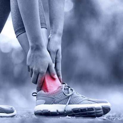
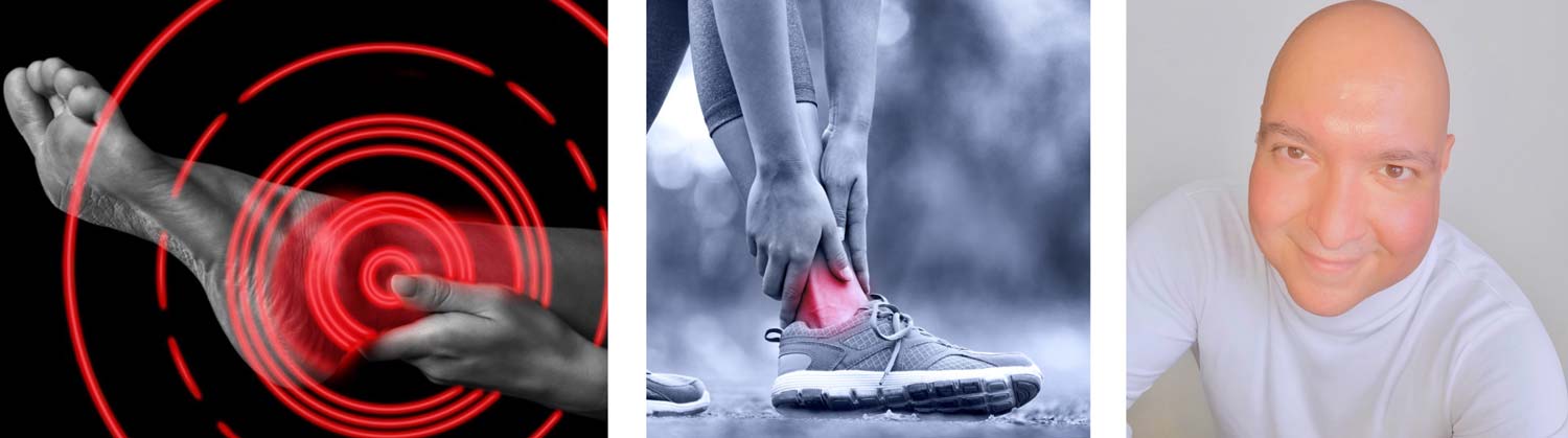
Consider the heel. When a runner wakes with knife-like pain at the plantar medial calcaneal tubercle that eases with gradual walking, the body is telling a story about the plantar fascia and its enthesis. The microtears of a stiff overnight fascia stretch across their weakest link as the windlass mechanism engages. A different patient, pointing slightly more lateral and distal with burning and paresthesias that worsen in narrow shoes, is describing something else: entrapment of the inferior calcaneal nerve (Baxter’s nerve) as it courses around the abductor hallucis and along the deep fascia. The 2 patients live in the same zip code; their “street addresses” differ by centimeters and by tissue type–collagenous fascia in 1, compressed nerve in the other. The treatment should differ just as precisely.
Pain mapping continues medially. Adult acquired flatfoot is not a generic “arch collapse.” It begins as degenerative change in the posterior tibial tendon, progresses to failure of the spring ligament, and eventually stresses the deltoid complex and the subtalar joint. Early pain is felt along the tendon’s distal course; as the deformity worsens, patients develop lateral impingement pain as the calcaneus everts and the lateral column is overloaded beneath the fibula. If one treats only the symptom at a single moment–an orthotic here, a steroid there–without naming where the cascade has moved anatomically, the plan will lag behind the disease.
On the lateral side, chronic ankle sprain is often anything but. Recurrent pain and a sense of giving way can reflect peroneus brevis split tears within a shallow retromalleolar groove, superior peroneal retinaculum injury with tendon subluxation, or sinus tarsi syndrome–synovial and ligamentous inflammation within the subtalar recess after inversion injury. These pathologies occupy discrete spaces. A patient who points behind the fibula and describes clicking with eversion is not asking for another ankle brace; they are asking the clinician to examine a tendon in a groove and a retinaculum under strain.
Pain Behavior is Biomechanics in Disguise
The timing of pain is as diagnostic as its location. Gait is an anatomic stress test that repeats thousands of times per day. Each phase highlights a different structure. At heel strike, the calcaneal fat pad, the cortical shell of the calcaneus, and the origin of the plantar fascia accept impact. If pain is maximal at initial contact, those structures move to the top of the list. Midstance demands controlled eversion and subtalar accommodation; posterior tibial tendon, spring ligament, and the midfoot joints carry the burden of stabilizing a flexible lever. Pain that builds through midstance signals failure of that support. Toe off loads the sesamoid apparatus, plantar plates, flexor hallucis longus, and Achilles tendon; pain with propulsion directs attention there. Pain that crescendos by day’s end reveals fatigue and overuse in tendons and ligaments; pain worst on first step after rest reflects tightening and enthesis irritation. The body is telling you when a given structure fails under load; it is your task to translate which structure that timing implicates.
Even quality of pain encodes anatomy. Burning, tingling, and electric shocks implicate nerves or their sheaths. Deep, dull ache that is worse after activity and improves with rest suggests bone or joint, particularly osteoarthritic surfaces sharing load poorly. Sharp, well-localized pain with stretch or palpation is often fascial or tendinous, especially at an enthesis. A sense of “catching” with particular arcs of motion implies mechanical conflict–an impingement spur, synovial fold, or loose body. Pain with passive versus active motion, pain with resisted testing, and pain with specific positions (forced dorsiflexion provoking anterior ankle impingement, forced plantarflexion awakening posterior impingement around an os trigonum) refine the map further.
The Clinical Error We Keep Making
The most common error in the care of foot and ankle pain is to convert the symptom into the label, then treat the label. “Plantar fasciitis” becomes a bucket that captures fascial enthesopathy, fat pad atrophy, calcaneal stress injury, and multiple neural entrapments in the distal tarsal tunnel complex. A steroid injection into the wrong bucket numbs the story for a few weeks while the real pathology continues. “Chronic ankle sprain” becomes a blanket term that hides peroneal tendon tears, occult fractures of the lateral process of the talus, and persistent synovitis in the sinus tarsi. “Bunion pain” is corrected with elegant osteotomies while first ray hypermobility, pes planus, or gastrocnemius equinus–the drivers of overload–go unaddressed, setting the stage for recurrence.
The antidote to this pattern is anatomical honesty. Pain is not the end of the history; pain is the beginning of a conversation with tissue. If we treat pain generically, it returns. If we treat the structure that is suffering and the mechanics that overburden it, the pain resolves because its reason for existing has been removed.
Two Stories That Change How We Listen
A 52-year-old nurse presents with 6 months of heel pain. It is worst with the first steps in the morning and after her 12-hour shifts. She has tried a night splint and an orthotic with minimal relief. Examination reveals focal tenderness 1–2 cm distal to the plantar medial calcaneal tubercle. The windlass test is positive; dorsiflexion of the hallux tensions the fascia and reproduces her pain. Ultrasound shows a thickened fascia at the enthesis with hypoechogenicity. This is the canonical fasciopathy: a collagen scaffold frayed by cumulative load. Now consider a second patient: a 46-year-old retail worker with plantar fasciitis for 9 months. She has burning pain that radiates to the plantar lateral heel, worsens in narrow shoes, and sometimes shoots forward into the lateral plantar foot. Tinel’s sign is positive at the abductor hallucis, and provocative eversion increases her symptoms. Ultrasound shows focal thickening of a small nerve branch deep to the fascia; the plantar fascia itself measures within normal range. Her pain was never fascial; it was neural. Both patients arrived with the same label. Only one was treated.
A second pair of patients illustrate the migration of pain with deformity. A 67-year-old man notes aching along the medial ankle and arch, worse by day’s end, with a new difficulty performing a single-limb heel rise. He has a valgus hindfoot, forefoot abduction, and a “too many toes” sign. MRI demonstrates degeneration within the posterior tibial tendon. Months later, as the tendon fails and the spring ligament attenuates, his pain moves to the lateral ankle with a sense of pinching beneath the fibula. The structure that hurts has changed because the alignment has changed; lateral impingement is a downstream consequence of medial collapse. Now consider a 28-year-old basketball player with recurrent “ankle sprains.” Despite bracing, he has posterolateral pain and a clicking sensation when he resists eversion. In clinic, the peroneal tendons subluxate with circumduction, and MRI confirms a split tear of the peroneus brevis within a shallow groove. His problem is not instability of the ankle joint; it is failure of a tendon and retinaculum in a tight space. The address matters.
Imaging as Confirmation, Not A Treasure Hunt
Imaging should follow anatomy; it should not try to find a diagnosis in the absence of an anatomic hypothesis. Ultrasound is unmatched for dynamic assessment of fascia, tendons, and superficial nerves; it is the clinician’s stethoscope for the plantar heel and peroneal tendons. It answers focused questions: How thick is the fascia at the calcaneal origin? Does the inferior calcaneal nerve change caliber as it exits the deep fascia? Do the peroneal tendons subluxate with active movement? Radiographs, especially weight-bearing views, reveal alignment and arthrosis in ways supine MRI cannot–subtalar alignment, talonavicular coverage, midfoot joint space narrowing, first ray elevation. MRI offers exquisite detail for osteochondral lesions of the talus, impingement syndromes, plantar plate tears, and the marrow response to chronic overload. But without a map drawn from history and examination, imaging becomes a scatter of findings in search of a story. Each modality is most valuable when it is deployed to confirm a structure you already suspect from the patient’s narrative.
How Nerves Write in Fire and Tendons Write in Ache
The language of pain varies by tissue. Nerves, trapped in fibro-osseous tunnels and sheathed in connective tissue that can swell, write in lightning and fire. They produce paresthesias, tingling, and numbness with a spread that follows their branching–posterior tibial nerve through the tarsal tunnel into medial and lateral plantar distributions; interdigital nerves beneath the deep transverse intermetatarsal ligament out into the workspace. Tendons write in ache that sharpens with stretch or resisted motion; their pain is focal at entheses and sweeping along sheaths if paratenon is involved. Ligaments write in instability and apprehension, especially at end-range motion that challenges their check-rein function. Bone writes in deep ache–stress reaction and arthritic surfaces that hurt after load and diffuse slowly.
Learning to hear those voices is as important as memorizing attachments. In practical terms, it means asking patients to describe not just “where,” but “what and when.” It means palpating in millimeters, not centimeters. It means provoking the structure you think is guilty–windlass for fascia, Tinel’s at the tarsal tunnel, single-limb heel rise for posterior tibial tendon, forced dorsiflexion for anterior impingement, plantarflexion for posterior impingement, resisted eversion for peroneals, drawer and tilt for ankle ligaments–and letting the structure answer.
The Role of Footwear, Surfaces, and Workload
Anatomy is not destiny; it is a set of tolerances that can be exceeded by environment and behavior. Footwear that narrows the forefoot compresses interdigital nerves; minimalist shoes expose calcaneal fat pads and plantar fascia to unfiltered impact; rigid high heels shift load to the metatarsal heads and sesamoids. Surfaces matter–concrete is less forgiving than track, treadmills alter stride, and cambered roads can torque ankles through repetitive valgus or varus control demands. Occupation shapes pathology. Nurses and retail workers report plantar heel pain not simply because they stand, but because they accumulate hours of midstance on unforgiving floors in footwear that may not address their foot posture. Runners exhibit predictable sequences of injury when weekly mileage outpaces tissue adaptation–Achilles insertional pain in the presence of a tight gastrocnemius, metatarsal stress reactions with sudden spikes in intensity, plantar fasciopathy during transitions to forefoot-strike patterns without graded strengthening of intrinsic muscles.
Misdiagnosis Patterns That Waste Time–and How to Avoid Them
Three patterns recur in clinics and deserve special attention. The first is “plantar fasciitis” that is not fascia. Baxter’s nerve entrapment, fat pad atrophy, and calcaneal stress injury can mimic it. Careful localization (more lateral and distal for nerve), provocative testing (Tinel’s at abductor hallucis), and ultrasound (nerve caliber, fascial thickness, fat pad integrity) separate them. Treating a nerve with a fascial injection is a recipe for delay.
The second is “chronic ankle sprain” that hides peroneal pathology or sinus tarsi syndrome. Patients who describe snapping or clicking behind the fibula with eversion, or who have pain to palpation in the sinus tarsi that feels like a deep bruise with inversion-eversion, need dynamic testing and targeted imaging. Tendon tears will not heal with more bracing, and sinus tarsi synovitis will not resolve with generic ankle rehab.
The third is bunion pain managed as if the first metatarsal were an isolated beam. First ray hypermobility, pes planus, and gastrocnemius equinus are common co-conspirators. If the ray drifts and the hindfoot collapses, the medial eminence is not the whole story; it is the visible symptom of an unstable construct. Osteotomies should be planned in a biomechanical context, and soft tissue balancing aims at restoring function, not merely image.
Why Anatomy Must Precede Intervention
Injections, orthoses, taping, physical therapy, shockwave, ablation, and surgery all have roles. None of them is a substitute for correct anatomical diagnosis. A custom orthotic that stabilizes a collapsing arch offloads the posterior tibial tendon and spring ligament; the same orthotic is irrelevant for a Morton’s neuroma and counterproductive for a patient whose pain is an impingement spur. A steroid into a fasciopathic enthesis can be carefully considered; a steroid into a nerve entrapment may quiet inflammation without relieving compression. Shockwave may stimulate healing in recalcitrant fascial degeneration; it is unlikely to help a split tendon. Ablating a neuroma addresses a choke point; failing to decompress the deep transverse intermetatarsal ligament leaves a mechanical trap in place.
Operative intervention, when necessary, is anatomy in its most literal sense. Arthroscopic debridement of anterior ankle impingement removes a physical block to dorsiflexion; os trigonum excision and FHL release clear a posterior conflict. Spring ligament augmentation, calcaneal osteotomy, and tendon transfer rebalance forces in adult acquired flatfoot; peroneal groove deepening and retinacular repair restore a pulley’s geometry. Cheilectomy in hallux rigidus relieves dorsal impingement and buys motion; fusion ends painful motion when cartilage is gone. Decisions are easy when the structure is named and hard when the label is vague.
Listening Like a Mapmaker
The clinicians who earn reputations for solving stubborn foot pain share an approach more than they share a trick. They listen for the time signature of pain, they palpate like cartographers, they test hypotheses at the bedside before they ever open the imaging viewer. They know that a positive windlass plus morning pain plus focal enthesis tenderness is fascia until proven otherwise; that paresthesias and Tinel’s with a more lateral point implicate a nerve; that posterior tibial tendon weakness will reveal itself when a patient tries to rise on tiptoe and the heel refuses to invert. They map pain onto anatomy and then confirm with targeted imaging. Their plans sound unglamorous: load management here, a different orthosis there, a decompression rather than a “clean-up,” an osteotomy to redirect force instead of an implant to mask symptoms. Their outcomes look like relief rather than magic.
A Brief Philosophical Detour: Pain as a Protective Truth
Pain has a bad reputation as the villain of clinical encounters. It is, in truth, a protective truth-telling mechanism. It forces a patient to stop before they tear what is only frayed; it pushes them to seek care before a nerve entrapment becomes irreversible damage; it motivates strengthening before a tendon surrenders in full. Our task is not to silence pain as quickly as possible; it is to understand the message and remove the cause. This attitude is not punitive; it is respectful. It treats the body as an intelligent system signaling its limits. Nowhere is that more evident than in the foot and ankle, where the economy of space and the volume of load leave little margin for error.
Pain is not mysterious, and it is rarely misleading. In the foot and ankle, pain tells the truth with an address. Our job is to read the street signs.
Dr. Hooman Mir, DPM, MSci, FAPWCA is a Tenure-Track Assistant Professor of Medicine and Faculty Senator at UTRGV School of Podiatric Medicine. Dr. Mir is an alumnus of Temple University School of Podiatric Medicine and completed his surgical internship at Mount Sinai Hospital in New York. Dr. Mir’s commitment to podiatric academic medicine is defined by a series of distinctive firsts: the first Doctor of Podiatric Medicine (DPM) to receive full NIH tuition reimbursement for a Master of Science in Clinical Investigation at UT Health San Antonio; the first DPM to graduate from Harvard Medical School’s prestigious T2T Program; the first DPM and only faculty member in the UTRGV Health System inducted into the historic Harvard Club of Boston; and the first DPM at UTRGV Health System ever to be accepted into the School of Medicine’s PhD program in Human Genetics–focused on Precision Medicine in Diabetes–now embarking on his second year of doctoral study.
- Balius R, Alomar X, Rodas G, Miguel-Pérez M, Pedret C, Vázquez R. The plantar fascia in sports players and sedentary population: Sonographic findings and correlation with clinical symptoms. Clinical Journal of Sport Medicine; 2013: 23(3), 210–215. doi: 10.1097/JSM.0b013e31827d0f07
- Baxter D, Pfeffer, G. Treatment of chronic heel pain by surgical release of the first branch of the lateral plantar nerve. Clinical Orthopaedics and Related Research;1992: 279, 229–236.
- Buchbinder, R. Plantar fasciitis. The New England Journal of Medicine; 2004: 350(21), 2159–2166. doi: 10.1056/NEJMcp032745
- Cheung J, Zhang M, An K. Effect of Achilles tendon loading on plantar fascia tension in the standing foot. Clinical Biomechanics; 2004: 19(8), 839–846. doi: org/10.1016/j.clinbiomech.2004.05.001
- Deland J, de Asla R, Sung I, Ernberg L, Potter H. Posterior tibial tendon insufficiency: Which ligaments are involved? Foot & Ankle International; 2005: 26(6), 427–435. doi: 10.1177/107110070502600604


