 While arthrodesis of the first metatarsophalangeal (MTP) joint is known to alleviate pain from osteoarthritis in the hallux, long-term outcomes vary, with nearly 20% of patients reporting dissatisfaction with the surgical outcome.
“There are a number of patients who have symptoms of pain, metatarsalgia, lateral toe arthritis or interphalangeal joint arthritis after 1st MTP joint arthrodesis,” said lead author Abbis H. Jaffri, from the Department of Kinesiology at the University of Virginia in Charlottesville. “However, the reasons for these problems post-surgery are not very well-known.” In the paper, he and 2 colleagues theorize that the post-surgical lack of motion in the 1st MTPJ results in compensation by lateral toes in an effort to restore foot function, which results in more loading on the lateral toes. This compensation can lead to osteoarthritis elsewhere in the foot and even metatarsalgia.
“These are the same problems that are experienced by a patient who has intrinsic foot muscle weakness,” Jaffri explained. “Therefore, we hypothesized that these issues experienced by patients following surgery may be due to the atrophy of these small muscles of the foot.” They devised an IRB-approved study using ultrasound (US) imaging to asses changes in the morphology and structure of the IFMs after 1st MPTJ arthrodesis. Their paper, “Ultrasound examination of intrinsic foot muscles in patients with 1st metatarsophalangeal joint arthrodesis,” was recently published in the journal The Foot.
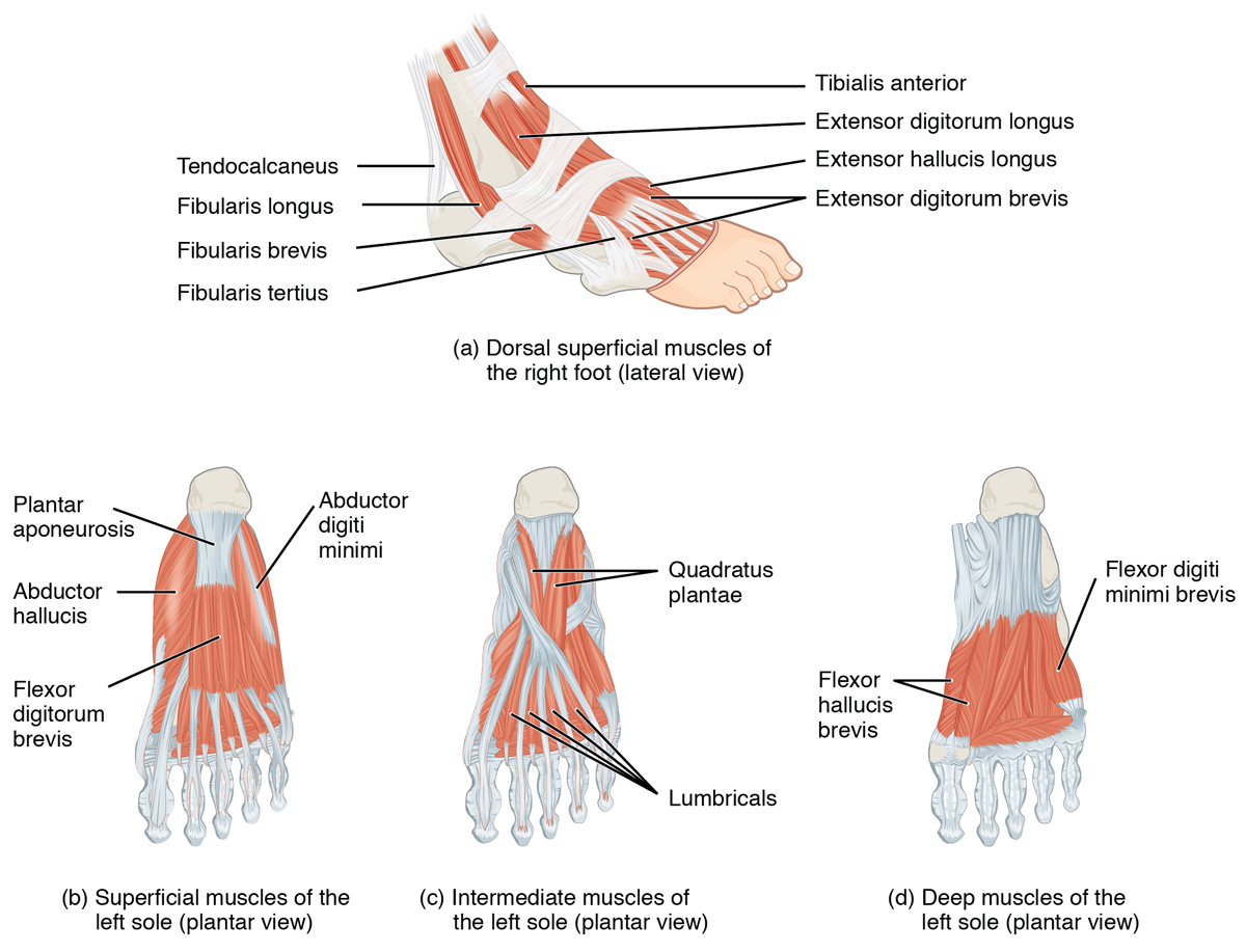
They recruited a convenience sample of 18 feet of 9 subjects with a unilateral history of 1st MTPJ arthrodesis; each participant served as their own control. Key demographics included: age: 57.56 ± 9.07; weight: 81.33 ± 1.32 kg; height: 163.26 ± 11.03 cm. B-mode ultrasound images of the cross-sectional area (CSA) and muscle thickness (MT) were collected of the Abductor Hallucis (AbH), Flexor Digitorum Brevis (FDB) and Flexor Hallucis Brevis (FHB) during sitting and standing positions with a wireless 8-MHz transducer. Toe flexion strength was measured with a hand-held dynamometer.
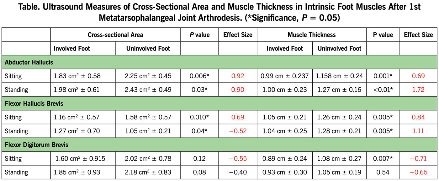 This paper delineates the atrophy of small yet significant muscles on the plantar surface of the foot in patients with 1st MTP joint Arthrodesis, said Jaffri (see Table). The authors also observed significantly lower (P < 0.05) toe strength in the involved toe compared to the uninvolved one.
The authors write that not only is this the first study to examine the plantar foot musculature after 1st MTPJ arthrodesis, but it is also the first time that IFMs have been assessed in a weight-bearing position. They note that FDB is the largest of the plantar IFMs and the observed atrophy could provide a potential explanation for the subsequent arthritis and metatarsalgia that some patients experience post-surgically. They further note that there are no protocols for rehabilitation after 1st MTPJ arthrodesis and that strengthening these muscles could help to improve functional outcomes after surgery.
“We did find significant atrophy of intrinsic foot muscles in this group of patients,” Jaffri said. “Identifying a problem is the first step in addressing it. This paper highlights a significant weakness that can be addressed through rehabilitation of foot and ankle post-surgery to improve functional outcomes.”
Source: Jaffri AH, Hertel J, Saliba S. Ultrasound examination of intrinsic foot muscles in patients with 1st metatarsophalangeal joint arthrodesis. Foot. 2019;41:79-84.


