
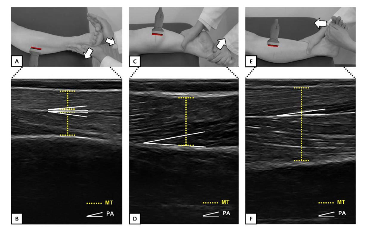
Figure 1. (A) Assessment of peroneal muscles at MVC; (B) ultrasound image of the peroneal muscles; (C) assessment of MGCM at MVC; (D) ultrasound image of the MGCM; (E) assessment of TA at MVC; (F) ultrasound image of the TA. MGCM, medial gastrocnemius; TA, tibialis anterior.
This study aimed to identify changes in the architecture and performance of the peri-ankle muscles in patients with chronic ankle instability (CAI) and investigate the relationship between them.
Inclusion criteria for the study included: (1) age ≥18 years or older; (2) previous history of ≥1 severe ankle sprain that caused pain, swelling, limited weight bearing, or complete immobility for ≥3 days; (3) failure to return to pre-injury functionality; (4) repeated episodes of ankle sprain; and (5) self-reported ankle dysfunction score of ≤24 on the Cumberland Ankle Instability Tool (CAIT), classified as a noticeable or pathologic condition. Exclusion criteria were as follows: (1) experienced an injury within the 3 months prior to first outpatient visit; (2) history of surgery on their bones, joint structures, or nerves in either lower limb; (3) previously experienced fracture in either lower limb; (4) acute injury to musculoskeletal structures in lower extremities within previous 3 months that impacted their joint function and resulted in ≥1 day of interrupted physical activity; (5) experienced current and/or intermittent pain; and (6) systemic musculoskeletal disease.
This retrospective cross-sectional study used data from the electronic medical records of patients with CAI who visited a tertiary care hospital during a 2-year period. In total, 17 subjects were evaluated retrospectively. Each subject underwent anthropometric and isokinetic test, and peroneus longus (PL) and brevis (PB), medial gastrocnemius (MGCM), and tibialis anterior (TA) ultrasound imaging were performed at rest and maximum voluntary contraction (MVC) conditions (Figure 1).
Regarding muscle architectural variables, the pennation angle (PA) of the MGCM at rest and the PA of the TA, MGCM, and PL in MVC were significantly reduced on the injured side compared to the intact side. There were no significant differences in muscle thickness of PL, PB, MGCM, and TA observed between intact and injured side during both rest and MVC.
Regarding muscle performance parameters, significant decreases were observed in the muscle strength for both limbs in all 4 directions under the 2 different conditions. A secondary finding was that the relative PA ratio of the TA showed moderate correlation with the relative dorsiflexion ratio at 30°/s.
These findings can provide opportunities to better understand how injuries in patients with CAI may be related to changes in ankle and foot function.
Source: Yu H, Yeo S, Lim JY, Kim I, Hwang J, Lee WH. Peri-ankle muscles architecture and performance changes in patients with chronic ankle instability: A retrospective cross-sectional study. J Foot Ankle Res. 2024;17(3):e12035. doi: 10.1002/jfa2.12035. Use is per Creative Commons License CC BY 4.0.


