 Research suggests that metatarsal pads and other off-loading techniques can help alleviate painful symptoms in patients with metatarsalgia. Orthotic intervention, in conjunction with correct shoes, can be an important component of improving quality of life for these patients.
by Paul R. Scherer, DPM
Multiple pathologies of mechanical origin that produce pain occur in the vicinity of the metatarsal heads, and these are lumped into a general category called metatarsalgia. Metatarsalgia is defined as pain from the area of five different metatarsal heads, five different metatarsophalangeal joints, and a variety of soft-issue structures in between.1 It usually occurs plantarly. Although commonly thought of as a symptom secondary to a more specific pathology, metatarsalgia is frequently categorized as both a symptom and a diagnosis.
A symptom of metatarsalgia is most often intractable pain that is difficult to resolve despite many attempts. The exact location of the pain is frequently elusive and often seems to migrate to different areas in the forefoot. The source of the pain must first be established before successful orthotic intervention can be attempted.
Primary metatarsalgia is defined as pain related directly to structure or function that results in a chronic imbalance of the pressure through the forefoot. Common diagnoses of primary metatarsalgia include second metatarsal stress syndrome, hallux valgus, brachymetatarsia, plantar flexed metatarsal, and iatrogenic symptoms secondary to a surgical procedure.1-7 Pain from primary metatarsalgia originates from a specific mechanical origin, and treatment should focus on accommodating or redirecting the specific mechanism.
Secondary metatarsalgia is considered pain related to a problem that does not originate within the metatarsal area.1 This “outside” force then produces problems or a mechanical imbalance affecting the metatarsophalangeal joint (MPJ) area. Typical etiologies for secondary metatarsalgia include rheumatoid arthritis, seronegative arthropathy, gout, infection, and equinus deformities. Treatment must be focused on both the systemic diagnosis and the local forefoot pain.
Metatarsalgia in the literature
A review of the published research related to metatarsalgia can help establish guidelines for clinical decision making.
A comprehensive study,1 published in 1980, found that 31 of 98 patients had two or more mechanical etiologies for their primary metatarsalgia, and that primary and secondary metatarsalgia are commonly found together in the same feet. This became a landmark study, since it exposed the complexity of metatarsalgia. Although many causes are possible, it seems that the mechanical origin component is common to both primary and secondary metatarsalgia, regardless of the original etiology. Even causes such as infection or iatrogenic postoperative problems ultimately result in a mechanical disruption of the metatarsophalangeal joint apparatus, and this mechanical dysfunction seems to produce the symptoms of metatarsalgia.
Further studies2-4 in patients with metatarsalgia and rheumatoid arthritis have focused on determining the level and source of biomechanical imbalance, whether it is from structure, gait pattern, disease process, or compensation. These studies suggest that focused evaluation with pressure plates, motion analysis, and injection therapy may help differentiate the source of the pain.
This approach may also aid in more directed and effective treatment with orthoses. Although not every case is complicated, it is useful to lay out a pathway to determine the treatments that may best contribute to a successful outcome for each patient. When a patient presents with pain and the compensatory signs of metatarsalgia, the importance of recognizing the cause cannot be overstated. Multilevel treatment of the originating cause, as well as the compensatory processes, must be considered. The success of orthotic therapy intervention is dependent on a thorough patient evaluation focused on the particular metatarsal region of the foot.
Orthotic therapy in the literature

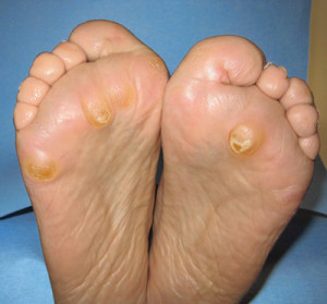
Figure 1. Persistent increase of forces for a prolonged time can dissipate the fat pad under the metatarsal heads and produce hyperkeratosis.
Reducing metatarsal head pressure has been the goal of treatment in several studies of orthotic intervention for metatarsalgia.5-8 The use of metatarsal pads in many shapes and sizes has been the most common method of in-shoe adjustment tested to relieve metatarsalgia symptoms. A metatarsal pad, placed behind the forefoot contact area, supports the metatarsal shafts and relieves pressure under one or all of the metatarsal heads.5 The more ground reaction force (GRF) that is shifted proximally to the metatarsal necks, the less force there is on the metatarsal heads.
A 1990 study used pedobarography to determine the effect of metatarsal pads on pressure under the metatarsal heads in 10 asymptomatic subjects.6 The effect of the intervention on the first metatarsal head was insignificant, although the pressure was significantly reduced at the second metatarsal head for all subjects. The pressure reduction from the use of the metatarsal pad decreased for each subsequent lesser metatarsal head lateral to the second metatarsal.
Another study of metatarsal pads, published in 1994, expanded on this idea by measuring the intervention’s effect on peak pressures in eight discrete plantar locations on the hindfoot, midfoot, and forefoot.7 In the 10 asymptomatic subjects studied, metatarsal pad use was associated with statistically significant increases in plantar pressure at the metatarsal shaft region, suggesting offloading from the metatarsal heads. Although there were no statistically significant changes in any other plantar region, there was a mild decrease in pressure at the first and second metatarsal heads and slight increases laterally. In addition, contact duration decreased at all metatarsal head locations and pressure-time integral (PTI) decreased at the first, second, third, and fourth metatarsals.
A number of important studies have assessed patients with complaints of metatarsalgia and their response to various treatments. The first study examined the difference between prefabricated orthotics and custom orthotics with and without rockerbars on the shoe.8 (Rockerbars are 6 mm to 10 mm thick strips of outsole material added just proximal to the metatarsal head portion of the shoe sole that are intended to increase GRF at the metatarsal shafts, therefore decreasing the force on the metatarsal heads.) In 42 patients with a history of metatarsalgia, the rockerbar in conjunction with prefabricated orthotics decreased pressure in the digital area of the forefoot, as did the custom orthotics with and without the rockerbar. Pain scores were significantly lower for the custom orthotics, both with and without the rockerbar.

Figure 2. Pressure mapping demonstrates that metatarsal pads can decrease peak pressures under the metatarsal heads.
Another intervention study was conducted on 33 subjects with complaints of metatarsal pain. Each subject used two different prefabricated soft insoles for a period of eight weeks.9 Plantar pressures were lower for both insole conditions relative to shoes without insoles in all subjects, and more than half of the patients reported relief of pain. There was no significant difference, however, between the two types of insoles. This study suggests that even unsophisticated intervention can have a positive effect on metatarsalgia.
A research group in Taiwan specifically evaluated the metatarsal pad to determine if the placement and location had an effect on peak plantar pressure reduction.10 A metatarsal pad measuring 55 mm in length, 36 mm in width, and 10 mm in height was uniformly used in 10 subjects with a history of primary metatarsalgia. Greatest pressure reduction was achieved when the metatarsal pad was placed just proximal to the site of peak metatarsal head pressure. The study revealed that positional differences as small as 4 mm could influence the metatarsal pad’s ability to reduce plantar pressures at the metatarsal heads. However, for clinicians to use the same type of assessment to place a metatarsal pad for each patient is impractical.
Since one of the most important outcomes of treatment is pain relief, in 2006 the same Taiwanese research group assessed the correlation between the use of a metatarsal pad and subjective symptoms.11 Thirteen patients with secondary metatarsalgia wore metatarsal pads under the second metatarsal head for two weeks. Improvements in visual analog pain scores were statistically correlated with reduction in pressure time integral and, more strongly, with reduction in maximum peak pressure.
Additional studies of secondary metatarsalgia have focused primarily on pain relief with the use of orthotics and metatarsal pads.12-14 These studies found that metatarsal pads were associated with statistically significant decreases in peak plantar pressures and pressure-time intervals and relieved patient pain and increased quality of life. The studies also found that the use of a custom orthosis without the pad provided a frequent decrease of peak pressures and decreased metatarsal pain in varying amounts.
The final step in learning how to treat a patient with metatarsalgia is to determine how to apply these data to an orthotic prescription for the patient.
A 2000 study of 24 subjects symptomatic for rheumatoid arthritis and MPJ pain compared the effectiveness of three interventions: supportive shoes alone, shoes with semirigid orthoses, and shoes with soft orthoses. Each intervention was tested for 12 weeks. A comparison of mean pain scores from baseline to final visit showed that semirigid orthoses significantly reduced pain, while the other two conditions did not.13
In a 2003 study, Mueller evaluated the primary forefoot structural factors that predict regional peak plantar pressure during walking in 20 people with diabetes mellitus and peripheral neuropathy and 20 controls. 15 The results showed that MPJ angle (hammertoe deformity) was the most important variable for predicting pressure in the diabetic group, accounting for 19% to 45% of the variance. Soft-tissue thickness, hallux valgus, and forefoot arthropathy were the most important predictors of PPP in the control group.
Although patients with diabetes rarely have metatarsalgia, the finding that structural MPJ deformity influences plantar pressure is interesting in light of other evidence suggesting that increased pressure at the metatarsal heads contributes to metatarsalgia and that the therapeutic reduction of pressure decreases symptoms of metatarsalgia.1,5,6,8,9 Studies of diabetic patients with ulcerations at metatarsal head locations have been useful in attempting to evaluate devices that decrease pressure in these areas. Although the two different pathologies are not necessarily related in terms of symptomatology, they may have similar pathomechanical origins.
In a 2006 study of 20 patients with diabetes, the same authors found that both a total contact insert and a metatarsal pad were associated with statistically significant reduction of pressure under the metatarsal heads.16 Subjects were analyzed while wearing just a shoe, a shoe with total contact orthoses, and a shoe with the same orthoses but with a metatarsal pad added. The total contact orthoses in this study increased the contact area with the foot by an average of 27%, primarily in the arch. The metatarsal pad, when added, did not provide any additional contact. The orthoses reduced metatarsal peak pressures by 19% to 24%. The PTI was reduced by 16% to 23%. The addition of a metatarsal pad, although it did not increase contact area, reduced peak pressure by an additional 15% to 20% and PTI by an additional 22% to 32%. The authors reported that the addition of a metatarsal pad increased the pressure peak at the second metatarsal shaft by an amazing 308%, indicating that although the pad did not increase surface area, it redistributed the metatarsal head pressure to the shaft.
Orthotic goals for metatarsalgia
The practitioner should start by establishing the etiology. Any systemic issues and subsequent foot changes due to compensation, as well as the mechanical origin of pain, must be established before determining the orthotic prescription. By creating a list of issues common to patients with primary or secondary metatarsalgia, the practitioner can develop an evidence-based orthotic prescription. The table that follows indicates the common biomechanical issues linked to metatarsalgia and the orthotic options that can be used with orthotic construction.
 Most of the studies that have been done regarding clinical intervention for metatarsalgia have involved the use of off-loading pads.1,5-12 The precise placement, according to the data discussed earlier, seems to be just proximal to the metatarsal head and just distal to the leading edge of the orthosis.10 The size and shape of the pads that are available vary, depending on the size and shape of the foot, as well as the metatarsals involved. There are, unfortunately, no data available on the most effective shape and size, and this lack of information may be the reason so many options exist. The use of metatarsal bars on the device, or on the shoe, also assists in relieving symptoms.
The orthotic plate should be made with minimal fill to create a total contact orthosis with the arch of the plate as high as the arch of the foot. This results in better contact of the orthotic plate against the plantar foot, increasing the intrinsic off-loading of the forefoot by the orthotic plate itself.
A study of 30 normal subjects confirmed that orthoses with minimum arch fill were far more effective than flat insoles for redistributing peak plantar pressures.17 The study demonstrated that the greater the contact in the arch area, the greater the decrease in rearfoot and forefoot pressures. The paper also revealed that the custom device made by either the CAD-CAM method or foam impression was far superior to the flat insole for reducing plantar pressures in the forefoot.
For every Newton of additional pressure on the arch area of the device, it seems logical that an equal Newton of pressure is taken away from the metatarsal heads. A semirigid or rigid device offers the “intrinsic off-loading” quality, since the force of the foot is fully supported by the orthotic plate. A flexible plate is not appropriate, since it compresses on loading, losing contact with the arch and increasing force at the metatarsal heads.

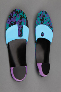
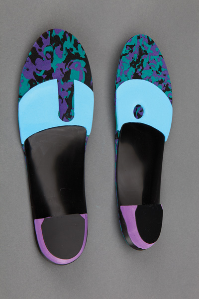
Figure 3. A combination of a forefoot foam addition, metatarsal bar, minimum positive arch fill, ethyl vinyl acetate (EVA) top cover, rearfoot post, and wide orthotic plate creates the recommended orthoses for metatarsalgia.
In my experience, a common component of overloading of the metatarsal heads is early heel-off. If the heel comes off the ground too quickly during the stance phase of gait, then logic suggests that the metatarsal heads in turn bear more weight for a longer period of time than they normally would. To compensate for this, a heel lift should be added on the orthotic post to bring the ground up to the foot, thus forcing the heel to maintain contact for a longer time with the ground before the propulsion phase of gait begins. The shifting of weight posteriorly reduces and delays pressure onto the forefoot.
Medial column instability due to excessive rearfoot pronatory motion often leads to dorsiflexion of the first ray and subsequent overloading of the second, limiting the first MPJ motion. A wide orthotic plate made from a cast with the first ray plantar flexed helps maintain contact with the more medial aspect of the foot, and it allows greater motion of the big toe and enhances weightbearing under the first metatarsal head, decreasing pressure under the second metatarsal head. 18
The body tissues lose their elasticity—and, therefore, their cushioning—with age. This process may also occur following surgery, after multiple injections, or with chronic, excessive pressure under the metatarsal head area. A top cover and a forefoot extension will add cushioning. Cushioning acts to decrease velocity, which dampens force. Research19 suggests that softer top‑cover materials give patients more comfort and increase their tolerance of orthotic devices, which may have implications for multiple pathologies. Materials such as closed-cell neoprene, plastazote, and Poron add shock absorption under the forefoot. Top covers require periodic replacement, dependent on patient weight, moisture, and frequency of use. Since compression of very soft materials over time negates the benefits of shock absorption that the patient needs, noncompliance can result if these materials are not replaced. Poron extensions that maintain their cushion ability under metatarsal heads also attenuate pressure by delaying compression.
Using the previously cited data from the literature, the goal of orthotic therapy is to reduce force on the metatarsal heads. The ideal functional orthosis for most metatarsalgia patients is a semirigid device made from a neutral suspension cast with the first ray plantar flexed. Cast correction should include a standard 14–mm heel cup, wide width, and minimum fill. The additions should include a rearfoot post with a small heel lift, metatarsal bar or pad, and soft, durable topcover with a soft, Poron extension.
Metatarsalgia is a common foot complaint. The onset is often slow, but the symptoms can be unrelenting and last a lifetime. Systemic disease and deformity contribute to severity. Using the variety of options and additions available on a custom orthotic, in conjunction with correct shoes, orthotic intervention can be an important component of improving quality of life and reducing pain in individuals with metatarsalgia.
Paul R. Scherer, DPM, is a clinical professor in the College of Podiatric Sciences at the Western University of Health Sciences in Pomona, CA. A version of this article appears in his book, Recent Advances in Orthotic Therapy, published by Lower Extremity Review. For more information, call 518/452-6898.
References
1. Scranton PE Jr. Metatarsalgia: diagnosis and treatment. J Bone Joint Surg Am 1980;62(5):723-732.
2. Tuna H, Birtane M, Tastekin N, Kokino S. Pedobarography and its relation to radiologic erosion scores in rheumatoid arthritis. Rheumatol Int 2005;26(1):42-47.
3. Woodburn J, Nelson KM, Siegel KL, et al. Multisegment foot motion during gait: proof of concept in rheumatoid arthritis. J Rheumatol 2004;31(10):1918-1927.
4. Laroche D, Pozzo T, Ornetti P, et al. Effects of loss of metatarsophalangeal joint mobility on gait in rheumatoid arthritis patients. Rheumatology 2006;45(4):435-440.
5. Valmassy R. Clinical Biomechanics of the Lower Extremities. St. Louis, MO: Mosby; 1996.
6. Holmes GB Jr, Timmerman L. A quantitative assessment of the effect of metatarsal pads on plantar pressures. Foot Ankle Int 1990;11(3):141-145.
7. Chang AH, Abu-Faraj ZU, Harris GF, et al. Multistep measurement of plantar pressure alterations using metatarsal pads. Foot Ankle Int 1994;15(12):654-660.
8. Postema K, Burm PE, Zande ME, Limbeek J. Primary metatarsalgia: the influence of a custom moulded insole and a rockerbar on plantar pressure. Prosthet Orthot Int 1998;2(1):35-44.
9. Kelly A, Winson I. Use of ready-made insoles in the treatment of lesser metatarsalgia: a prospective randomized controlled trial. Foot Ankle Int 1998:19(4):217-220.
10. Hsi WL, Kang JH, Lee XX. Optimum position of metatarsal pad in metatarsalgia for pressure relief. Am J Phys Med Rehabil 2005;84(7):514-520.
11. Kang JH, Chen MD, Chen SC, Hsi WL. Correlations between subjective treatment responses and plantar pressure parameters of metatarsal pad treatment in metatarsalgia patients: a prospective study. BMC Musculoskelet Disord 2006;7:95.
12. Hodge MC, Bach TM, Carter GM. Orthotic management of plantar pressure and pain in rheumatoid arthritis. Clin Biomech 1999;14(8):567-575.
13. Chalmers AC, Busby C, Goyert J, et al. Metatarsalgia and rheumatoid arthritis—a randomized, single blind, sequential trial comparing two types of foot orthoses and supportive shoes. J Rheumatol 2000;27(7):1643-1647.
14. de P Magalhaes E, Davitt M, Filho DJ, et al. The effect of foot orthoses in rheumatoid arthritis. Rheumatology 2006;45(4):449-453.
15. Mueller MJ, Hastings M, Commean PK, et al. Forefoot structural predictors of plantar pressures during walking in people with diabetes and peripheral neuropathy. J Biomech 2003;36(7):1009-1017.
16. Mueller MJ, Lott DJ, Hastings MK, et al. Efficacy and mechanism of orthotic devices to unload metatarsal heads in people with diabetes and a history of plantar ulcers. Phys Ther 2006;86(6):833-842.
17. Ki SW, Leung AK, Li AN. Comparison of plantar pressure distribution patterns between foot orthoses provided by the CAD-CAM and foam impression methods. Prosthet Orthot Int 2008;32(3):356-362.
18. Scherer PR, Sanders J, Eldredge, DE, et al. Effect of functional foot orthoses on first metatarsophalangeal joint dorsiflexion in stance and gait. J Am Podiatr Med Assoc 2006;96(6):474-481.
19. Pawelka S, Kopf A, Zwick E, et al. Comparison of two insole materials using subjective parameters and pedobarography. Clin Biomech 1997;12(3):S6-S7.


it is very informative and educative for who working in podiatry field