








 By Tom Chang, DPM
1. The Foundational Philosophy: The Proximal-to-Distal Algorithmic Approach
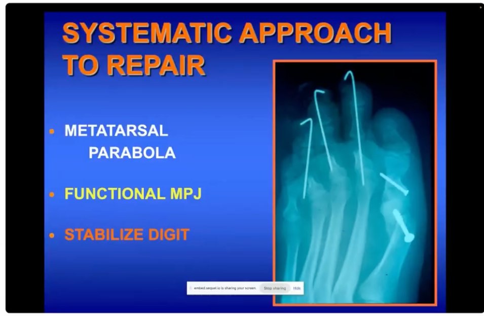
In the demanding arena of revision digital surgery, success is predicated on a strict “Proximal-to-Distal” hierarchy. The primary driver of surgical failure in this discipline is the isolated digital approach–addressing the toe as a localized deformity without considering the global biomechanical forces acting upon it. Long-term stability requires the neutralization of these forces through a systematic stabilization of the forefoot foundation. By establishing a sound proximal construct and then progressing distally, the surgeon ensures that the distal reconstruction is protected from the recurrent deforming stresses that compromised the index procedure.
The evolution of these principles emerged from the 1990s McGlamry/Atlanta era, a period defined by a high volume of complex referrals for severe digital complications. During this era, early iterations of Minimally Invasive Surgery (MIS) often utilized non-fixated osteotomies, allowing metatarsals to “find their own level.” This lack of structural control frequently resulted in unpredictable alignment and mechanical failure. Modern MIS has since evolved into an anatomically grounded discipline, yet the lessons of that era underscore our current reliance on rigid fixation and anatomical realignment.
To achieve predictable outcomes, the reconstructive algorithm must prioritize three tiers:
- Restoration of the Metatarsal Parabola: Re-establishing functional length and weight-bearing patterns.
- Metatarsophalangeal Joint (MPJ) Balancing: Correcting articular orientation and soft tissue tension.
- Digital Stability: Final stabilization of the digit via arthrodesis or tendon balancing.
The metatarsal parabola is the non-negotiable starting point for all revision work. Without a balanced weight-bearing construct, any distal repair is anatomically predisposed to recurrence.
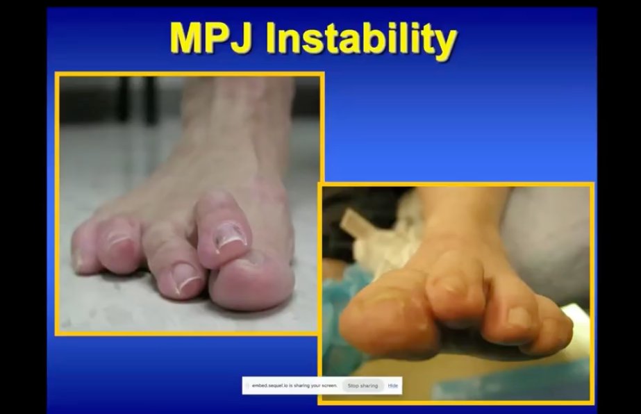 2. Tier I: Restoration of the Metatarsal Parabola
The metatarsal parabola serves as the engine of functional weight-bearing. When this length pattern is disrupted, the forefoot cannot distribute pressure predictably, leading to localized overloading. Failure to restore this parabola ensures that structural stresses will continue to cause secondary digital deviation or “floating” toes. Shortening and decompression osteotomies are essential tools for re-establishing these patterns and offloading high-pressure areas.
Diagnostic criteria for these interventions must begin with an assessment of the medial column. An unstable first ray or a significant hallux valgus deformity is often the primary culprit behind lesser MPJ stress, as the lack of medial support leads to massive transfer loading. Stabilizing the first ray is frequently a prerequisite for protecting the second and third rays. Whereas 1990s-era non-fixated MIS often failed due to a lack of structural control, modern principles dictate that the metatarsals must be anatomically positioned and fixated to provide a stable platform for the MPJ.
3. Tier II: MPJ Realignment and Soft Tissue Balancing
The MPJ is the functional “hinge” of the forefoot. Revision surgery requires meticulous balancing of both the articular cartilage orientation and the surrounding soft tissue tension. A critical clinical pearl is the evaluation of the metatarsal head cartilage: if the joint surface is oriented plantar, or medially or laterally, the toe will never remain rectus post-operatively, as the joint will naturally drift back to its malaligned orientation once temporary pins are removed.
The surgical protocol for MPJ realignment follows a strict sequential release algorithm:
- Step 1: Extensor lengthening
- Step 2: Dorsal, medial, and lateral capsulotomy.
- Step 3: Flexor plate release.
Validation: A Kelikian push-up test must be performed after each stage of release to clinically validate that the soft tissue tension has been neutralized.
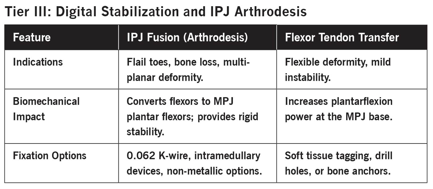 If the articular cartilage is malaligned, the surgeon could employ a “mini-Akin” phalangeal osteotomy or a metatarsal head wedge osteotomy to physically redirect the joint surface. In salvage scenarios where extra support is required, syndactyly to an adjacent digit offers excellent local stability. For chronic dislocations with redundant soft tissue and distal migration of the fat pad, an “elliptical plantar skin resection” is helpful to remove the excess tissue mass that would otherwise act as a deforming force post-operatively. Once the MPJ is secure, digital-level procedures may proceed.
4. Tier III: Digital Stabilization and IPJ Arthrodesis
Digital stability is achieved by converting the flexor tendons from deforming forces into active plantar flexors of the MPJ. In revision settings, Interphalangeal Joint (IPJ) fusion is the preferred method for providing long-term structural rigidity, especially when dealing with previous arthroplasty failures.
Surgical handling of tenuous revision tissues requires skin-to-bone target dissection. By maintaining full-thickness flaps and avoiding traditional anatomic layer dissection, the surgeon preserves the marginal circulation of the scarred digit, minimizing the risk of vascular compromise.
 5. Specialized Technique: Autogenous Calcaneal Bone Grafting for Flail Toes
The flail toe results from aggressive bone resection or severe instability, presenting a significant reconstructive challenge. To restore length and structural rigidity, autogenous bone from the lateral calcaneus is the gold standard due to its superior integration. Allograft options are available, but the autograft from the heel is my preference.
Step-by-Step Procedural Guide:
Harvest: Through a lateral approach to the calcaneus, harvest a unicortical rectangular segment of bone. Ensure the graft contains both cortical and cancellous bone for optimal strength.
The “Shish-Kebab” Technique: To prevent graft fragmentation, do not use a large wire initially. First, pass a 0.035 or 0.045 K-wire centrally through the graft and the digital bones.
Fixation: Once the central pin placement is confirmed, replace the pilot wire with a 0.062 K-wire for definitive stability. The wire should be driven through the distal tip of the toe, through the graft, and retrograde across the MPJ for tri-plantar stability.
Postoperative Protocol: Protection of the pin is required for 8 weeks to allow for early consolidation. While the result may be a stable fibrous union (pseudoarthrosis) rather than perfect bony bridging, the restoration of length and stiffness provides a successful clinical outcome.
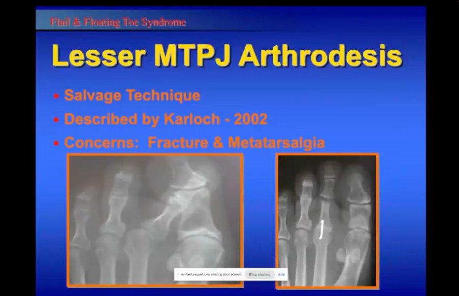 6. Advanced Salvage: Pan-Metatarsal Head Resection and Complex Deformity
In “train wreck” scenarios, where three or more rays are compromised, attempting to salvage individual joints often results in cumulative failure. In these cases, the rationale is to perform a biomechanical reset via a pan-metatarsal head resection.
Partial Metatarsal Head Resection: Indicated for 1–2 compromised rays, particularly in older populations. This requires 6 weeks of pinning and is remarkably successful for sagittal plane realignment.
Pan-Metatarsal Head Resection: Reserved for “3-out-of-5” ray involvement. While this provides a predictable reset for sagittal plane deformities, it is less effective for transverse plane deviations, which may require more aggressive soft tissue work.
Medial Anchoring: A concomitant first MPJ fusion is highly recommended during pan-resections. This provides a stable medial anchor for the reconstructed parabola and prevents the medial column from drifting.
While cosmetic outcomes in salvage cases may be compromised by scarring or shortening, the restoration of structural parameters remains the primary metric of success. By adhering to the proximal-to-distal algorithm, the surgeon can convert complex digital failures into a functional and stable foot.
Dr. Thomas J. Chang is a board-certified and highly skilled podiatric surgeon with over 30+ years of experience treating a wide range of foot and ankle conditions. He is currently a Professor and co-chair of the department of podiatric surgery at Samuel Merritt University College of Podiatric Medicine. He is also faculty of The Podiatry Institute.
Dr. Chang graduated with his doctorate degree from the Temple University School of Podiatric Medicine, where he was a member of the Pi Delta National Podiatry Honor Society and recipient of the Professor Louis M. Newman Surgery Award. He then completed a three-year residency program in Podiatric Medicine and Surgery at Emory/Northlake Regional Medical Center.
To further hone his skills and knowledge, Dr. Chang completed additional training at workshops and seminars across the country, focusing on ankle arthroscopy, cartilage repair, foot and ankle fixation and microsurgery. He also provided numerous lectures for Continuing Medical Education courses to impart his expertise to other doctors.
In addition to his training and practice duties, Dr. Chang also holds several appointments at local hospitals and medical schools, including:
California College of Podiatric Medicine–clinical professor & past chairman of Department of Podiatric Surgery
Health South Surgery Center–Director of Surgical Residency Training
Santa Rosa Memorial Hospital–Chief of Podiatric Medicine and Surgery
This article is a summary of Dr. Chang’s presentation, “Complications of Digital Surgery” from the 2026 No-Nonsense Seminar, March 6-8. To view the full presentation with questions and answers–and see the agenda for the program, visit https://nononsense2026.lerexpo.com/. Continuing education credits are available for this and many of the lerEXPO programs.
Related posts:
The Future of Bike Fit (Part 2) – Interview of the Bike Fit Machine Manufacturers and Bike Fit Education Content Creator...

Surgery Versus Watchful Waiting for First Metatarsophalangeal Joint Osteoarthritis
AI Coach Assists with Exercise Form, Provides Guidance

Examining Effect of Tai Chi Lower Leg Exercise in Fall Prevention in Older Adults

Machine Learning Combined with Explainable Artificial Intelligence Facilitate Accurate Balance Ability Classification