








 By Dr. Dennis Janisse
In the quiet halls of the Gillis W. Long Hansen’s Disease Center in Carville, Louisiana, the legendary Dr. Paul Brand once faced a clinical mystery.
He was conducting a study with a hypothesis that seemed like a mathematical certainty: total bed rest should heal a foot ulcer faster than any other intervention. By removing the variable of weight-bearing pressure entirely, the skin should have had the perfect environment to repair itself.
But the data began to fail. Results were inconsistent, and wounds that should have been closing remained stubbornly open. The breakthrough didn’t happen in a lab; it happened during a midnight walk through the infirmary. As Dr. Brand moved through the darkened wards, he discovered the culprit: the “bed rest” patients were getting up to use the bathroom unprotected.
Those few steps—taken without the “orthopedic jail” of a Total Contact Cast (TCC) to provide absolute immobilization—were enough to negate weeks of progress. This realization fundamentally changed our understanding of diabetic foot care. It proved that even minimal movement can be catastrophic if we ignore the horizontal forces acting on the skin. To truly save the diabetic foot, we must look beyond the squeeze of vertical pressure and address the silent, internal trauma of shear.
 Friction is a Rub, but Shear is Scissors
In clinical practice, “friction” and “shear” are often used as synonyms but treating them as interchangeable is a mechanical mistake. Friction is the surface-level resistance to motion between two bodies—it is the “rub” you feel when you slide your hands together. Shear, however, is far more insidious. It is the internal force that occurs when the skin stays gripped to a surface while the underlying skeleton continues to move.
If friction is the rub, shear is the scissors. It is the force that “clips” or “trims” the internal tissue layers, leading to the mechanical breakdown that precedes blisters and ulcers.
The danger lies in the relationship between the two: shear happens because the friction at the surface is too high. When the skin is “stuck” to an insole or a sock, the internal tissues are stretched and distorted as the bone moves forward during gait. In a patient with loss of protective sensation, these scissor-like forces cause significant internal damage long before an open wound ever appears.
The “Hole” Fallacy and the Danger of Window Edema
When a “hot spot” or ulcer appears, the instinctual, yet flawed, reflex is to “donut” the wound—cutting a hole in the orthotic to provide relief. While this technically removes vertical pressure from the center of the wound, it creates a phenomenon known as “window edema.”
Because there is a total lack of counter-pressure within the void of the hole, the unsupported skin quickly fills with fluid (edema). This creates a destructive cycle:
- The skin herniates into the hole, becoming swollen and engorged.
- The edges of the hole create new, high-pressure borders against the swollen tissue.
- As the patient walks, this fluid-filled “window” of skin rubs intensely against the sharp margins of the relief.
Instead of providing a sanctuary for healing, the hole becomes a site of intense friction and shear at the margins, often enlarging the wound or preventing the edges from closing.
 The Plastazote Paradox
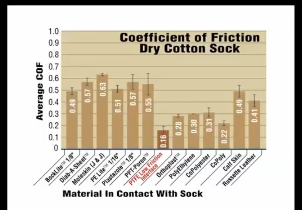
In the world of pedorthics, Plastazote #1 is often considered the gold standard. It is beloved for its ability to provide a dynamic floor reaction imprint. Because it compresses easily, it acts as a diagnostic tool; when a patient returns for a follow-up, the clinician can see exactly where the foot is bottoming out.
However, Plastazote presents a paradox: it is soft, but it is sticky at a microscopic level.
- Plastazote coefficient of friction: 0.57
- Low-friction interface materials: 0.16
This is where the “Plastazote Paradox” can endanger a patient. A soft material that grips the skin can be more damaging than a harder material that allows it to slide. Because Plastazote has such a high coefficient of friction, it holds the skin in place while the bones of the foot move inside, creating the very shear forces we are trying to avoid.
The “Mailman’s Secret”: The Power of 2 Socks
One of the most effective ways to manage the “pistoning” and “twisting” forces of walking is a surprisingly low-tech solution. There was a case of a mailman who continued to deliver mail for years on a professional route—even after undergoing amputations of his second metatarsal and great toe. His secret? He always wore 2 pairs of socks.
The logic is a masterclass in sacrificial mechanics:
- By wearing 2 layers, the friction is sacrificed at the interface between the 2 socks.
- The inner sock stays stationary against the skin, while the outer sock moves with the shoe.
- The rub occurs between fabric layers rather than against living tissue.
This simple strategy mimics high-end medical interfaces by ensuring that the shear forces are absorbed by the hosiery, sparing the skin from internal tearing.
Proactive Protection with Low-Friction Interfaces
We should not wait for the skin to break to act. Proactive management involves identifying “hot spots”—areas of red, warm skin—and applying low-friction interfaces before an ulcer forms.
A breakthrough in this area was developed by Marty Carlson in Minneapolis: a material known as ShearBan. This low-friction interface is a proactive tool that allows the foot to glide during high-impact movements. For clinicians, it includes a built-in “wear indicator”: the material is blue on top and white underneath. When the blue layer wears away to reveal the white, it is a clear clinical signal that the material needs replacement.

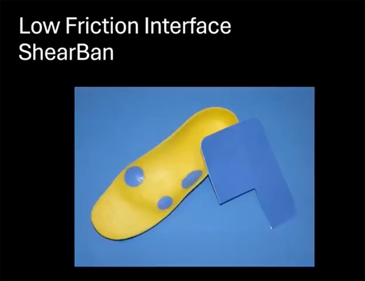
Low-friction interfaces are transformative across various pathologies:
- The Athlete: A basketball player with chronic, game-ending blisters can find immediate relief by covering the orthotic with a low-friction interface, allowing the foot to glide during high-impact pivots.
- The RA Patient: Patients with rheumatoid arthritis and claw toes often suffer from painful calluses at the tips of the digits. By placing a low-friction material under the toes, the calluses often disappear—even if the toes continue to “claw” during gait—because the rub component has been removed.
- The Diabetic/Charcot Foot: For patients with scar tissue (which lacks the mobility of healthy skin) or bony prominences, adding a low-friction layer proactively prevents the re-ulceration that often follows the buildup of a protective callus.
“Better wisdom is red skin equals a rub area or friction area as well and that leads to more options and better solutions.”
Moving Toward a 2-Vector Approach
To advance the standard of care for the diabetic foot, we must adopt a 2-vector approach to biomechanics. We can no longer afford to focus solely on the vertical vector of pressure while ignoring the horizontal vector of shear.
While offloading pressure remains a pillar of treatment, it is only half the battle. By managing the friction at the interface—whether through Marty Carlson’s specialized materials, the mailman’s double-socking method, or by avoiding the donut hole fallacy—we provide the skin the stable, low-shear environment it needs to remain intact.
If we are only measuring pressure, what invisible forces are we allowing to undermine our healing?
Dennis Janisse, C.Ped of National Pedorthic Services, Inc., a full-service Pedorthic facility. In addition to his duties at NPS, Mr. Janisse is a Clinical Assistant Professor in the Department of Physical Medicine and Rehabilitation at the Medical College of Wisconsin. He teaches pedorthics courses at Northwestern University Medical School, Century College and the Medical College of Wisconsin. He is past President of the Pedorthic Footwear Association and an honorary member of the American Orthopaedic Foot and Ankle Society. He has written extensively on all aspects of Pedorthics, with over 50 publications to his credit and has lectured throughout the United States and the world.
Related posts:

Consumer Exoskeleton for Hiking, Running

Ewing Sarcoma of The Great Toe: A Rare Case Report
lerEXPO Highlights: Digital Solutions for Early Detection, Prevention and Personalized Treatment of the Diabetic Foot
Wedge Fixation System for Evans and Cotton Osteotomies
lerEXPO Highlights: Fibular Nails–Fad or New Standard?