







 By Amiethab Aiyer, MD
1. Evolution and Biomechanical Foundations of MIS Hallux MP Arthrodesis
The strategic transition from open to minimally invasive surgery (MIS) for hallux metatarsophalangeal (MTP) arthrodesis represents a significant shift in delivery, yet the biomechanical mandate remains unchanged. Correction mechanisms in the forefoot are fundamentally grounded in soft-tissue dynamics. Regardless of the surgical approach, the realignment of the hallux is predicated on the appropriate tensioning and pull of the surrounding structures.
In the context of the stiff/arthritis hallux valgus deformity, the adductor complex serves as the workhorse for deformity correction. By leveraging the adductor’s pull, the surgeon can effectively narrow the intermetatarsal (IM) angle. This correction occurs at the joint level, but its success is dictated by these proximal soft-tissue tensions.
Biomechanical data comparing fixation methods reveal that the traditional preference for plate-and-screw constructs is not necessarily superior in an MIS environment. Studies evaluating arthroscopic joint debridement followed by percutaneous screw fixation demonstrate that screw-only constructs provide greater stiffness and equivalent resistance to plantar gapping when compared to plate-and-screw alternatives. This theoretical stability, however, is only realized through rigorous technical execution; without it, the clinical reality of fusion rates quickly diverges from these biomechanical ideals.
2. Clinical Literature Review: Benchmarking Success and Failure
For the surgeon, establishing clinical Is critical for identifying preventable failures. Understanding the standard complication rate allows the consultant to differentiate between biological outliers and technical suboptimization.
The following table summarizes current benchmarks for MIS hallux arthrodesis:
A critical finding in the literature is the 2% incidence of interphalangeal (IP) joint arthritis. An audit of these cases suggests this is not a random biological event but is likely related to the malpositioning of the toe during the index procedure. Furthermore, the asymptomatic non union remains a specific post-operative risk, appearing in approximately 6% of cases. While these patients may remain functional, the absence of radiographic fusion underscores the thin margin for error. These benchmarks serve as a reminder that successful outcomes are inextricably linked to the technical imperatives of joint preparation.
3. Technical Imperatives: The Anatomy of Joint Preparation and Fixation
 Joint preparation is the foundational pillar of MIS arthrodesis; any compromise here renders the subsequent fixation irrelevant. The surgical auditor must insist on a protocol that ensures the complete removal of articular cartilage and the exposure of subchondral bone.
Technical execution demands the use of the 2 x 12 mm Shannon burr. However, the use of high-speed burrs introduces the risk of thermal necrosis. Proper Thermal Management, specifically monitoring burr temperature and using intermittent irrigation, is mandatory to prevent bone death at the fusion interface. Regarding visualization, if arthroscopic assistance is not utilized, the surgeon must use intensive fluoroscopic visualization of the entirety of the joint or transition to a mini-open approach to confirm resection, just beyond the subchondral plate.
Technical Gold Standards for Fixation
To achieve a stable mechanical environment, the following “Gold Standards” must be strictly adhered to:
- Sagittal Plane Positioning: Perform the “pulp of the finger” check to verify appropriate extension. This check is vital to ensure the toe is not too straight (leading to tip-of-toe pressure) or too extended (causing dorsal pressure issues).
- Screw Spread: Achieve maximum multi planar stability through a wide, divergent distribution of screws across the joint.
- Bicortical Purchase: Verify on fluoroscopy that all screws achieve purchase in both cortices to optimize compression.
- Rotational Control: Use derotational control wires to prevent the spinning of the phalangeal segment during screw insertion.
A departure from these standards is not a variation in technique but a technical suboptimization that predictably leads to failure.
4. Failure Mode Analysis I: Technical Suboptimization and Delayed Union
 Lack of cortical purchase in the proximal phalanx can be a reason for delays in healing; this may be an important factor in healing of an MIS hallux MP fusion.
In failed constructs, this phalangeal screw is often oriented too medially, failing to bridge the width of the metatarsal head and missing the lateral cortex entirely. This lack of lateral purchase, combined with inadequate screw spread, leaves the joint vulnerable to the biomechanical stresses of weight bearing. While these mechanical failures may be technically related,, they can be exacerbated by poor biological management.
5. Failure Mode Analysis II: The Crux of the Problem in Material Reactions
In a recent audit of a chronic pain patient, a silica-based synthetic bone graft was utilized. This choice became the primary driver of a catastrophic failure. The silica graft instigated an excessive inflammatory response, presenting clinically as extreme, recalcitrant swelling. This inflammatory milieu likely contributed to the development of a Methicillin-sensitive Staphylococcus aureus (MSSA) colony. The resulting complications, including the infection and severe post-operative nausea/vomiting, led to an extended and unnecessary hospital stay.
Computed tomography (CT) imaging revealed the hallmark signs of this biological failure:
- Massive joint diastasis and resorption of bone surrounding the synthetic material
- Clear loosening of all fixation screws
- A robust inflammatory reaction that effectively dissolved the fusion site
The subsequent revision, which required the removal of all synthetic material and the application of stable fixation with autograft, resulted in successful consolidation. This case proves that material choice can override technical precision; no amount of screw torque can compensate for an inflammatory environment that actively promotes bone resorption.
6. Litigating Future Decisions: A Framework for Risk Mitigation
 Refining surgical judgment requires the surgeon to litigate every case, identifying where technical and biological choices diverged from the ideal. The following checklist provides a framework for mitigating future risk:
- Joint Prep Integrity: Resection must be verified beyond the subchondral bone across the 3D geometry of the joint.
- Thermal Management: Maintain constant vigilance of burr temperature to prevent thermal necrosis.
- Implant Selection: Weigh the necessity of staples or plates against crossing-screws based on bone quality.
- Biological Strategy: If necessary, consider autograft for biologic augmentation.
- Cortical Purchase: Mandate fluoroscopic verification of bicortical stability for every screw.
MIS hallux arthrodesis is a highly viable procedure that offers patients reduced pain and faster recovery. However, these benefits are only attainable when the surgeon operates within a strict framework of technical and biological rigor.
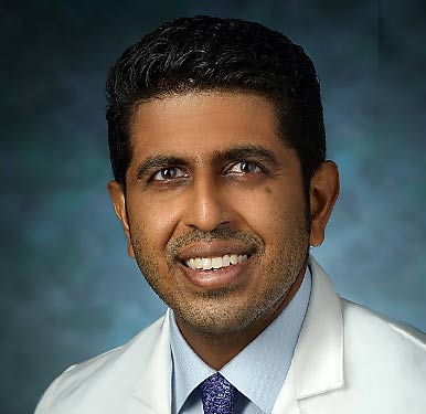
Amiethab Aiyer, M.D., is an orthopaedic surgeon specializing in treating patients with foot and ankle conditions, such as fractures, deformities, and ligament and tendon tears. He is skilled in minimally invasive bunion correction, Achilles tendon rupture repair, as well as ankle reconstruction and replacement. Dr. Aiyer is the division chief of foot and ankle surgery in the Johns Hopkins Department of Orthopaedic Surgery. He completed his medical training at Drexel University College of Medicine in Philadelphia. During his fourth year of medical school, Dr. Aiyer completed a research fellowship at the Rothman Orthopaedic Institute, also in Philadelphia. He then completed an orthopaedic surgery residency at the Penn State Health Milton S. Hershey Medical Center in Hershey, Pennsylvania. During the residency, Dr. Aiyer received multiple awards, including the Alan Levine Orthopaedic Graduate Medical Education Scholarship from the American Academy of Orthopaedic Surgeons (AAOS). He has also been selected to present at the Orthopaedic Research and Education Foundation Resident Research Symposium and as an American Orthopaedic Association (AOA) Emerging Leader. In his final year of residency, he received the Gold Humanism Resident Teacher Award. Dr. Aiyer went on to complete a research and clinical fellowship at the Institute for Foot and Ankle Reconstruction at Mercy Medical Center in Baltimore. As a researcher, he is currently investigating diagnosis, surgical treatments and patient outcomes for bunions, Achilles tendon ruptures, ankle arthritis, ankle trauma and other conditions affecting the foot and ankle. His research encompasses clinical, basic science and biomechanical studies. He has presented his research at numerous conferences held by the American Orthopaedic Foot & Ankle Society, AAOS and other professional organizations of which he is a member. He is a fellow of the AAOS and AOA, and a member of the Alpha Omega Alpha Honor Medical Society. Along with special interests in clinical practice and multiple facets of orthopaedic foot and ankle research, he is passionate about teaching residents and medical students; he has been mentoring medical students for several years. Dr. Aiyer has been recognized for his commitment to education with multiple teaching and faculty educator awards, and he is working with other orthopaedic surgeons to develop a nationwide push for medical student mentorship. Using social media platforms, he co-founded the @orthomentor Instagram account to provide students with easy access to mentorship and advising.
This article is a summary of Dr. Aiyer’s presentation, “MIS Forefoot Complications Session: Case 1” from the lerEXPO MIS Complications Expo December 6 , 2025. To view the full presentation with questions and answers—and see the agenda for the program, visit https://mis4.lerexpo.com/. Continuing education credits are available for this and many of the lerEXPO programs.
Related posts:
Benefits of Buerger-Allen Exercises for People with Diabetes: A Mini-Review

Therapeutic Shoes Work for People with Diabetes. Why Doesn’t Every Patient Use Them?
Shape-Shifting Scaffold Fights Infection, Rebuilds Bone

Injuries Related to Sauna Bathing
Technology Is Inevitably Reinventing Orthotics and Prosthetics