CONFERENCE COVERAGE: Orthotics Technology Forum 2013
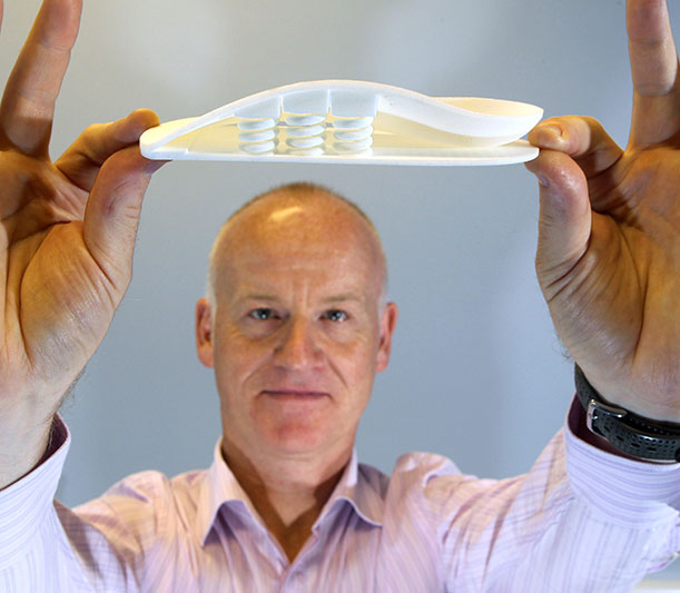
An image of the future shown at the 2013 OTF: A 3D-printed model of the human foot, part of the A–FOOTPRINT project (see “3D printing,” page 40). (Photo courtesy of Peter Devlin.)
By Emily Delzell
One of the most-talked-about images from the 2013 Orthotics Technology Forum (OTF) depicted 11 custom foot orthoses made by 11 experienced orthotists and podiatrists for a single patient. All 11 practitioners had been given the same information about the patient’s condition, yet all 11 orthoses were distinctly different (see figure, page 40).
The image, part of a presentation by Nachiappan Chockalingam, MSc, PhD, CEng, CSci, underscored the opportunity for advanced technology to take the guesswork out of orthotic prescription—a theme that was repeated throughout the OTF, which was held in late May at the Georgia Institute of Technology in Atlanta.
More than 80 attendees enjoyed several days of presentations, conversation, and a tour of Georgia Tech’s clinical biomechanics labs and the associated Global Center for Medical Innovation. In addition to host university Georgia Tech, event sponsors included Delcam Healthcare Solutions, Freedom Machine Tool, nora systems, SureFit, Mile High Orthotics Lab, Walking Mobility Clinics, Acor, Kiwi, and Kintec.
Drilling down into foot function
Chockalingam, professor of clinical biomechanics at Staffordshire University in Stoke-on-Trent in the UK, and others in his lab are combining plantar pressure and imaging data to develop patient-specific modeling to make orthotic prescription more accurate.
“[We can] provide more detail about foot function through mathematical models that could keep things like this from happening,” he said, pointing to the image of the 11 different orthoses.

Differences in orthotic training and approach result in highly variable prescriptions using raditional methods. (Image courtesy of Nachiappan Chockalingam, MSc, PhD, CEng, CSci.)
His team looks at both static and dynamic measurements using motion-capture systems and marker sets; force plates; in-shoe and mat plantar pressure systems; and systems that use interdigital sensors to measure dynamic plantar pressure. They also use ultrasound and magnetic resonance imaging to look at changes in foot tissue under different conditions.
“We also use multisegmental foot models to measure relationships between different foot segments rather than measuring simple angles and range of motion,” he said. “For example, we’ll look at the relationship between the medial forefoot and the lateral forefoot, the forefoot and the rearfoot, the rearfoot and the shank.”
Chockalingam and his colleagues have developed a device that combines ultrasound with dynamometry to measure mechanical response to plantar loading. His team developed this as part of its involvement with DiaBSmart (development of a new generation of diabetic footwear using an integrated approach and smart materials), an international effort funded by the EU.
They found normal pressure differential gradients were 6.25 times higher in patients with diabetes than in controls, a factor that may be an independent predictor in diabetic foot ulceration, Chockalingam said.
“Physiological changes can affect mechanics, and various technologies can help us develop patient-specific and predictive models that can alter the orthotic prescription and process,” he said.
A closer look at tissue behavior, stiffness
In his Georgia Tech lab, research scientist Géza F. Kogler, PhD, CO, is also combining imaging and pressure measurement tools to produce unique data on tissue behavior and stiffness characteristics of the foot.
“There are all these different soft tissues through which you’re having to transfer a load [when an orthosis is used], and if I want to transfer a load I need to know what those tissues are and what their different compositions and mechanical properties are,” he said in an OTF presentation.
Kogler, who is director of Georgia Tech’s Clinical Biomechanics Laboratory, and his team have developed test apparatuses to quantify plantar midfoot soft tissue stiffness properties and behavior in nonweight-bearing and weight-bearing conditions to provide perspective on the diversity of tissues.
“We had a fairly simple hypothesis; that plantar midfoot tissues would be stiffer in weight bearing than in nonweight bearing because, when we load the foot, its structures become tense,” he said. “Most tissue studies have been done at the heel, but most of the control in an arch mechanism is at the midfoot, so we started there.”
They took measurements from a halfway point between the metatarsal heads and the posterior aspect of the calcaneus under weight-bearing and nonweight-bearing conditions, using ultrasound to quantify tissue displacement.

“We proved our intuitive hypothesis, that tissues were stiffer under weight-bearing than nonweight-bearing conditions, and also saw that tissue behavior and stiffness varies in individuals due to BMI and individual differences in tissue structure,” said Kogler, who had tested 25 healthy volunteers at the time of the OTF (the study is ongoing).
“What we’re really after is the clinical application [of this research], and to get to where we can determine what that stiffness value is in a particular foot and how we might use that to our advantage to control the movement with an orthosis, which means we need to alter the shape and perhaps the density of the material,” he said.
Kogler also discussed pilot study data that showed an orthosis changed the stiffness properties of plantar tissues compared with a barefoot condition.
“We’ve used a rigid device that changes shapes and manipulates stiffness, and also changed the alignment of this person’s foot structure from pes planus to one that has a slight arch. So, as orthotists, we have huge control capabilities—we can control skeletal structure and stiffness by changing shape, and we may be able to make similar changes by changing materials.”


