
iStockphoto.com #697961234
Poor vision has long been known as a risk factor for poor balance. This is particularly true in older adults as they perform complex balancing tasks, such as standing on 1 foot.
A team of Canadian researchers wanted to determine whether poor vision would be more strongly related to standing balance in older adults who had peripheral vascular disease (PVD) or diabetes. Using information from the Canadian Longitudinal Study on Aging, researchers recruited 30,097 adults (45 to 85 years of age) who lived near 1 of the 11 data-collection sites in 7 Canadian provinces. Data were collected between 2010 and 2015. using various questionnaires that were administered either by an interviewer at home or at a data-collection site. Peripheral vascular disease and diabetes status were assessed according to self-reporting of a physician’s diagnosis.
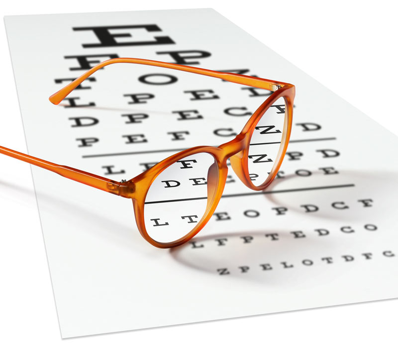
Physical exams were performed at the data-collection sites by trained members of the research team, who used the Early Treatment Diabetic Retinopathy Study chart at a 2-meter distance to measure participants’ visual acuity while they were wearing habitual distance correction.
Researchers then used the “1-leg standing test” to assess participants’ balance. This test can predict falls and disability that falls can cause. Only participants who could stand unassisted were asked to perform the balance test. Without shoes, participants stood approximately 3 feet from a wall and were instructed to balance on 1 foot while lifting the other leg to calf level with their hands on their hips. Participants were allowed to practice the procedure before the timed test. Poor balance was defined as being unable to stand on 1 leg for at least 60 seconds.
Participants who did not attempt the balance test were compared to those who did. The findings show that people with poorer vision were more likely to have balance problems. Other factors related to poor balance included:
- peripheral vascular disease
- being female
- low level of education
- being a current smoker
- higher body mass index (BMI)
- type 1 or type 2 diabetes.
Visual acuity and PVD were indeed linked: The odds of having poor vision were almost double in people who also had PVD, compared to those who did not have it (odds ratio [OR] = 1.50; 95% confidence interval [CI], 1.29–1.77). In the group without PVD, after adjusting for age, sex, education, province, BMI, and diabetes status, a 1-line worse score on the visual acuity test was associated with 23% higher odds of not being able to stand for the full 60 seconds (OR = 1.23; 95% CI, 1.20–1.26). In the group with PVD, the odds of not being able to stand was nearly doubled (OR = 1.41; 95% CI, 1.22–1.62).
Although the researchers found that the interaction between visual acuity and PVD was statistically significant (P = .02), they also determined that the interaction between diabetes and visual acuity was not significant (P = .11). They concluded by suggesting that patients with known PVD should be encouraged to see an optometrist or ophthalmologist for a vision test. Likewise, patients with known balance challenges should have an ankle-brachial determination to rule out PVD.
Vafaei A , Aubin MJ , Buhrmann R , Kergoat MJ , Aljied R , Freeman EE. Interaction between visual acuity and peripheral vascular disease with balance. J Am Geriatr Soc. 2018;66(10):1934-1939.


