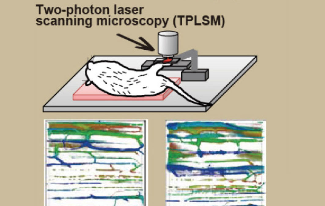
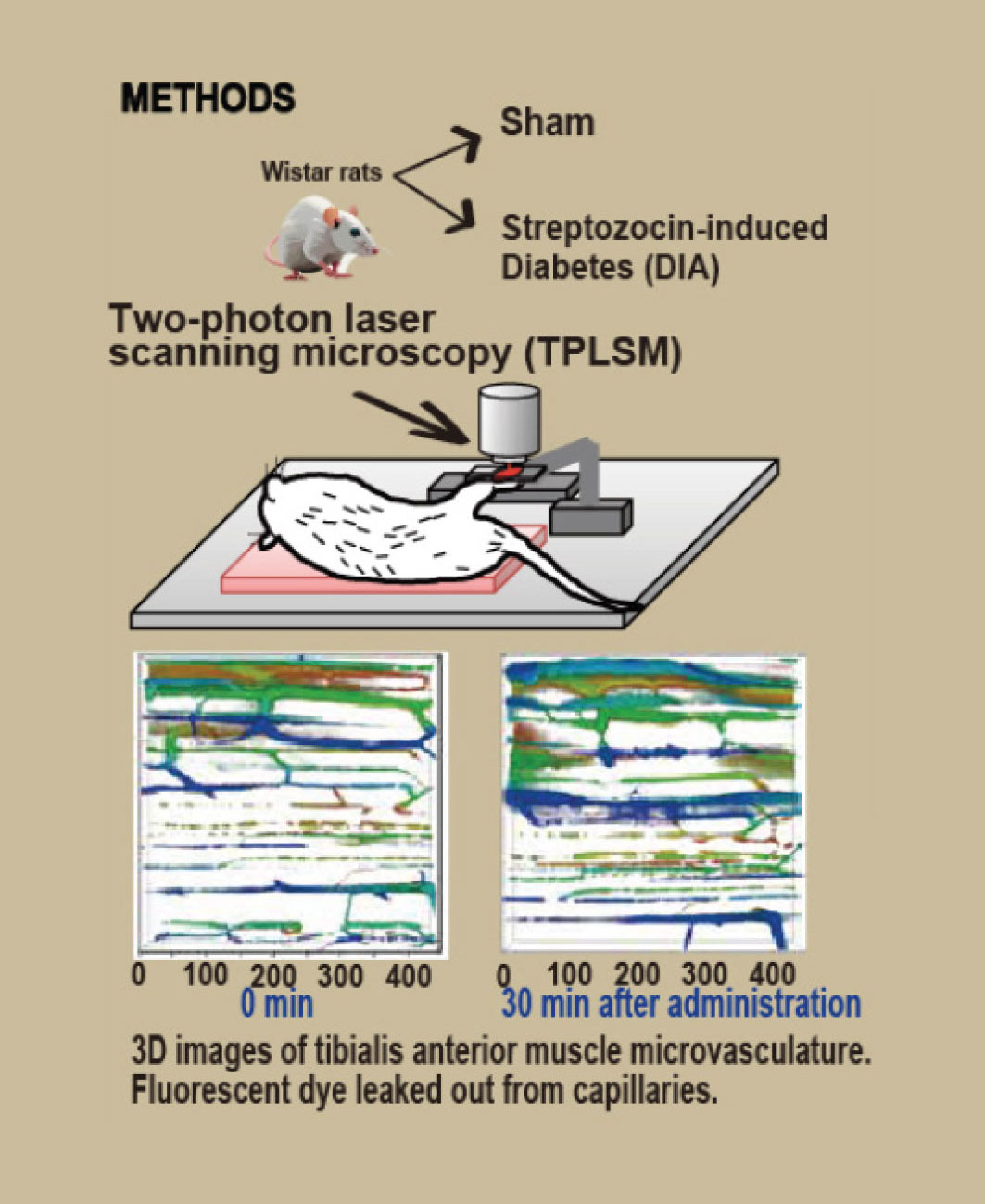
 Microvascular permeability in diabetic muscle was investigated using our original 2-photon scanning laser microscopy method. Compared with controls, the leakage volume was increased in diabetic muscle, which was atrophic with smaller capillary diameter, endothelial cell thickening, and the appearance of more endothelial intercellular gaps or clefts, and large vesicles. Hyperpermeability was closely related to ultrafine structural changes of the capillary endothelial cell junctions.
Source: Hotta K, Shimotsu R, Behnke BJ, et al. Effect of diabetes on microvascular morphology and permeability of rat skeletal muscle: in vivo imaging using two-photon laser scanning microscopy. J Appl Physiol (1985). 2024;137(4):963-974. doi: 10.1152/japplphysiol.00222.2024.


