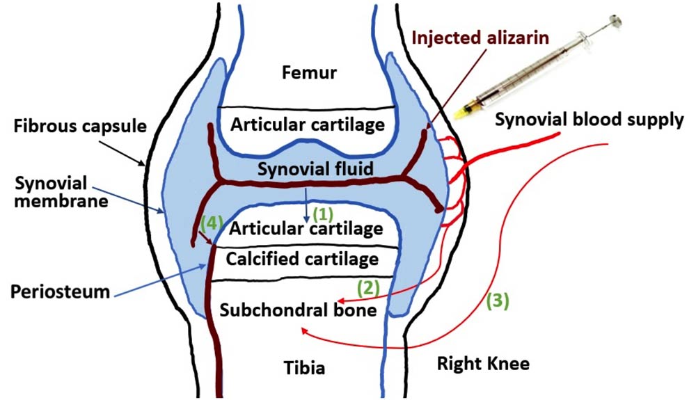
The fourth diffusion route is a novel diffusion direction showing that alizarin complexone can pass into the periosteum in the marginal transitional zone of the tidemark.
A fluorescent dye could help scientists listen in on biochemical conversations between cartilage and bone during the earliest stages of osteoarthritis (OA)—even before the disease causes pain. The unexpected finding could someday lead to novel treatments for patients, according to research conducted on mice.
Bin Wang and colleagues at the Sidney Kimmel Medical College of Thomas Jefferson University wanted to know whether articular cartilage becomes calcified early in OA, so they studied a mouse model in which the right knee exhibits symptoms similar to the human disease. A fluorescent red dye called alizarin complexone binds calcium-containing crystals, and the researchers injected this dye into both knees. Surprisingly, there was no fluorescence staining on the surface of the articular cartilage layer—where the team expected to find new calcification—in early OA stages.
The tidemark area, a barrier between the articular cartilage and a layer of calcified cartilage that resides on the bone was stained in both knees. “But we found more of the alizarin dye in the calcified cartilage and subchondral bone in osteoarthritic mice compared to control,” said Wang. Increased diffusion of the dye suggests that the early-OA knee joint is more permeable than the control.
With an injection, dye first goes directly into the synovial fluid that cushions the joints. In additional experiments, the researchers found that the dye could then move throughout the joint via 3 expected pathways. However, they also observed a fourth, brand-new pathway into the blood vessels in the outer covering of the bone, called the periosteum, via the tidemark. The fluorescence signal was greater in the periosteum, as well as in the subchondral bone of early-OA mouse joints than in controls.


