“This is the first prosthetic study in history that shows a leg prosthesis under full neural modulation, where a biomimetic gait emerges,” said Herr. Image courtesy of Hugh Herr and Hyungeun Song.
Using a new type of surgical intervention and neuroprosthetic interface, Massachusetts Institute of Technology (MIT) researchers, in collaboration with colleagues from Brigham and Women’s Hospital, have shown that a natural walking gait is achievable using a prosthetic leg fully driven by the body’s own nervous system. The surgical amputation procedure, known as the agonist-antagonist myoneural interface (AMI), reconnects muscles in the residual limb, which allows patients to receive proprioceptive feedback about where their prosthetic limb is in space.
Most limb movement is controlled by pairs of muscles that take turns stretching and contracting. During a traditional transtibial amputation, the interactions of these paired muscles are disrupted, making it very difficult for the nervous system to sense the position of a muscle and how fast it’s contracting—sensory information that is critical for the brain to decide how to move the limb. People with this kind of amputation may have trouble controlling their prosthetic limb because they can’t accurately sense where the limb is in space. Instead, they rely on robotic controllers built into the prosthetic limb. These limbs also include sensors that can detect and adjust to slopes and obstacles.
To try to help people achieve a natural gait under full nervous system control, Hugh Herr, PhD, a professor of media arts and sciences, co-director of the K. Lisa Yang Center for Bionics at MIT, and an associate member of MIT’s McGovern Institute for Brain Research, and his colleagues began developing the AMI surgery several years ago. Instead of severing natural agonist-antagonist muscle interactions, they connect the 2 ends of the muscles so that they still dynamically communicate with each other within the residual limb. This surgery can be done during a primary amputation, or the muscles can be reconnected after the initial amputation as part of a revision procedure.
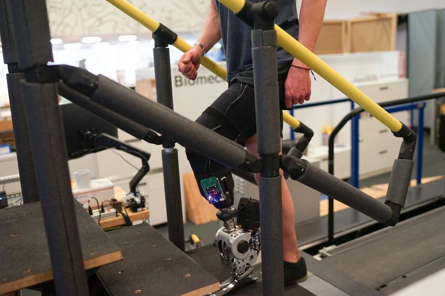
For this study, the researchers compared 7 people who had the AMI surgery with 7 who had traditional transtibial amputations. All of the subjects used the same type of prosthesis with a powered ankle as well as electrodes that can sense electromyography signals from the tibialis anterior the gastrocnemius muscles. These signals are fed into a robotic controller that helps the prosthesis calculate how much to bend the ankle, how much torque to apply, or how much power to deliver. The researchers tested the subjects in several different situations: level-ground walking across a 10-meter pathway, walking up a slope, walking down a ramp, walking up and down stairs, and walking on a level surface while avoiding obstacles.
In all of these tasks, the people with the AMI neuroprosthetic interface were able to walk faster—at about the same rate as people without amputations—and navigate around obstacles more easily. They showed more natural movements, such as pointing the toes of the prosthesis upward while going up stairs or stepping over an obstacle, and they were better able to coordinate the movements of their prosthetic limb and their intact limb. They were also able to push off the ground with the same amount of force as someone without an amputation. These natural behaviors emerged even though the amount of sensory feedback provided by the AMI was less than 20% of what would normally be received in people without an amputation. They also experienced less pain.


