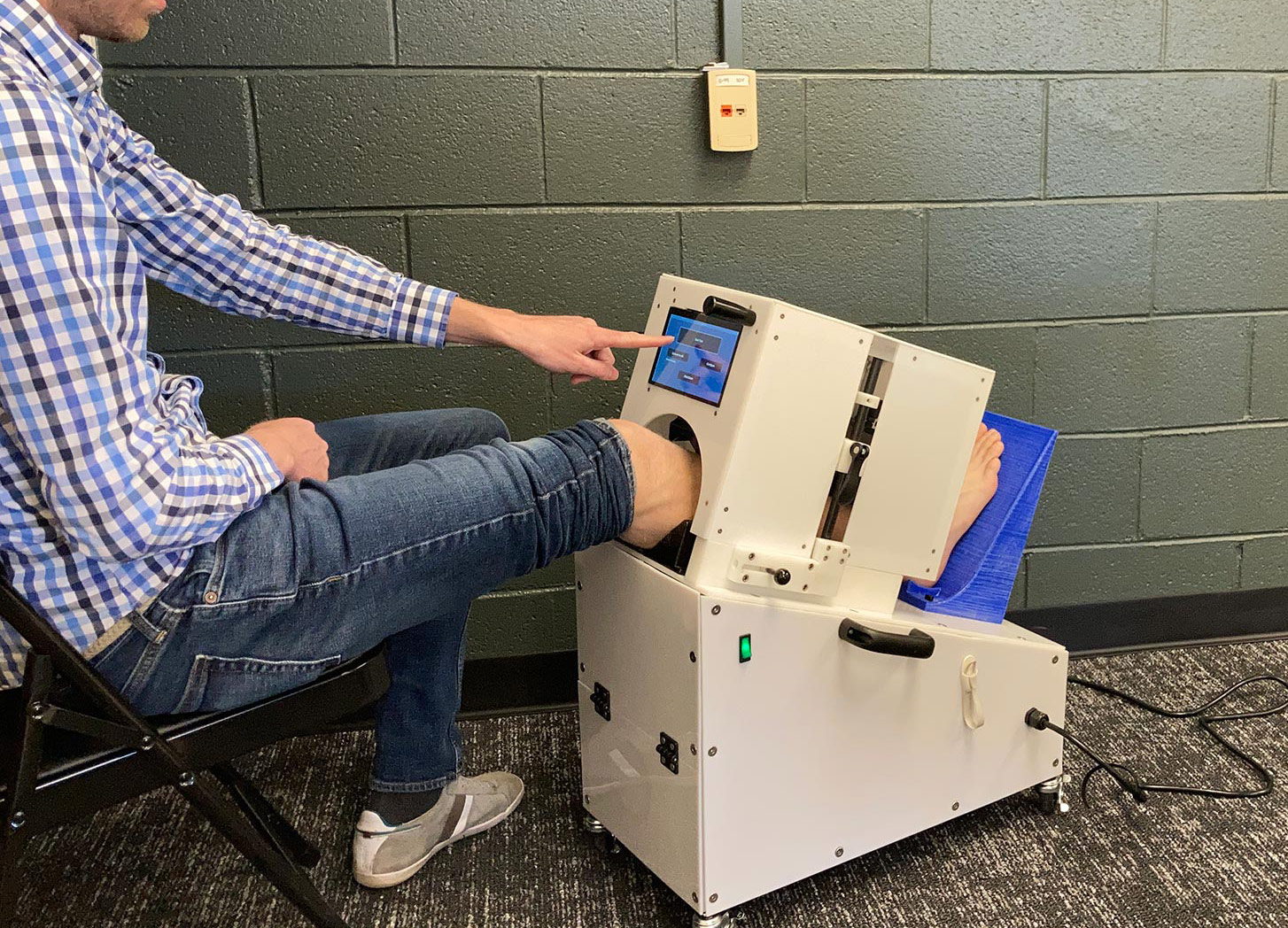
CSU research scientist Kevin Labus demonstrates the tibia fracture device, now being used in a study with patients.
People who fracture their tibia usually get hardware surgically implanted to hold the bone in place. After about 6 months, 10% of patients are diagnosed with a non-union fracture; they require additional surgery to implant new hardware. The standard method to diagnose a non-union fracture is through x-ray, but x-rays cannot capture the subtle mechanical details of how well bone tissue is mineralizing and stiffening soon after the injury. What if there was a way this stiffening could be detected much earlier than 6 months post-injury, so doctors could intervene faster?
Nearly 2 decades ago, Colorado State University biomedical researcher Christian Puttlitz first put his mind to this question, wondering whether innovations in engineering and biomechanics could provide insight into the healing potential of bone. From that inspiration sparked a series of experiments that led to a present partnership with UC Health doctors and patients, who are currently helping test a fracture-detection device Puttlitz and his lab have been working on for the last several years.
The device consists of an enclosure that applies gentle, non-painful pressure on the patient’s fracture site. An external radio antenna sensor measures deflection under loading, and the sensor is calibrated to measure bending stiffness of the fracture callus. Down to 10-micron precision—with much more accuracy and detail than an x-ray—the device lets the researchers assign a value to how well the bone is stiffening—an indication of normal healing—starting about 6 weeks after the initial injury. That’s much sooner than the 6-month mark patients usually have to wait to find out whether their fracture is mending properly.