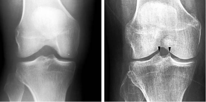
AI tries to detect whether there is spiking on the tibial tubercles in the knee joint, which can be a sign of OA. Image courtesy of University of Jyväskylä.
Researchers from the University of Jyväskylä, Finland, and the Central Finland Health Care District have developed an artificial intelligence (AI)–based neural network to detect early knee osteoarthritis (OA) from x-ray images. The new AI-based method was trained to detect from x-rays whether there is spiking on the tibial tubercles in the knee joint or not. The finding is not at the moment included in the diagnostic criteria, but orthopedic specialists consider it as an early sign of OA.
“Around 700 x-ray images were used in developing the AI model, after which the model was validated with around 200 x-ray images,” said Anri Patron, the researcher responsible for the development of the method. “The model managed to make an estimate of the spiking that was congruent with a doctor’s estimate in 87% of the cases, which is a promising result.”
The result is important because x-rays are the primary diagnostic method for early knee OA, and an early diagnosis can save the patient from unnecessary examinations, treatments, and even knee joint replacement surgery. In addition, the patient might be motivated to take the measures to slow down or even stop the progression of the symptomatic OA.
The goal is that in the future, AI would be able to detect early signs of knee OA from x-rays, making it possible for the initial diagnosis to be made more often by general practitioners.