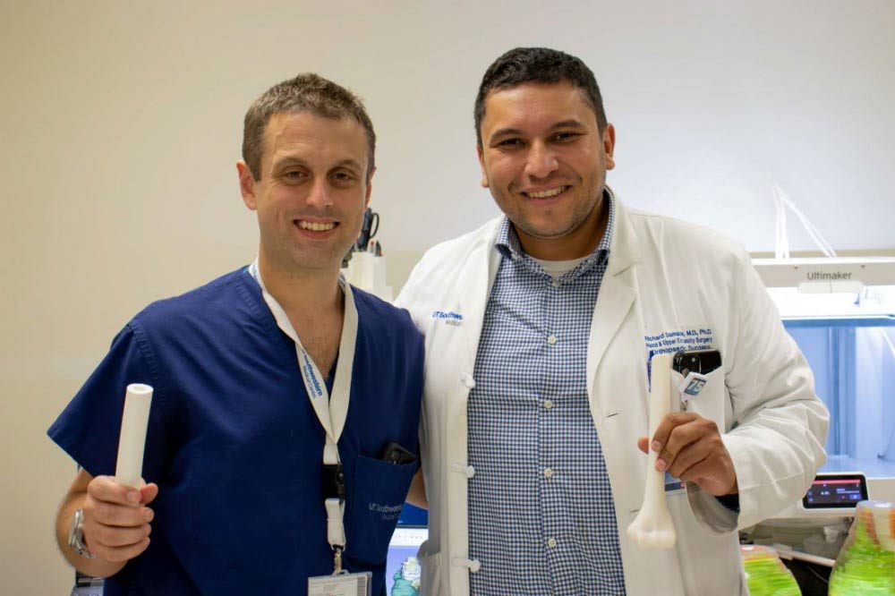
Weinschenk (left) and Richard Samade, MD, PhD, assistant professor of orthopedic surgery, biomedical engineering, and plastic surgery at UT Southwestern Medical Center, collaborated with mechanical engineers from UT Dallas on the research.
Researchers at University of Texas (UT) Southwestern Medical Center have developed a breakthrough three-dimensional (3D) printing technique for generating realistic models of the human femur that could make it easier and less expensive to conduct biomechanical research. Though the work focused on replicating the femur and its unique mechanical properties, the process could be used in the future to build models of any human bone for research.
Collaborating with mechanical engineers from UT Dallas, Robert Weinschenk, MD, assistant professor of orthopedic surgery and biomedical engineering at UT Southwestern, and the team used polylactic acid—an inexpensive, biodegradable polyester material commonly used in 3D printing—to construct a wide range of femur models with different physical attributes such as wall thickness and infill density. Those models were then tested for flexural strength using 3-point bending, and the results were compared to the biomechanical response of human femurs, enabling the team to identify the methodology that produced the most accurate replica.
Researchers at UT Dallas focused on the mechanical evaluation and characterization of the 3D-printed femur. “With 3D printing, we’re able to print out the femur bone with the same geometry of the femur inside the body,” said Wei Li, PhD, assistant professor of mechanical engineering at UT Dallas. “In our biomechanical tests, the femur performed as well as a human femur.”


