 Researchers from the UK have identified three subgroups of individuals with patellofemoral pain (PFP) that can be determined using simple evidence-based clinical tests—a key first step toward developing targeted treatment strategies as part of the ongoing effort to improve PFP outcomes.
By Jessie Janssen, PhD; James Selfe, DSc; Michael Callaghan, PhD; Chris Sutton, PhD; Paola Dey, MD; and Jim Richards, PhD
In recent years the patellofemoral pain (PFP) research community has highlighted the need to identify subgroups of patients with PFP, with the aim of being able to develop specific interventions tailored for each subgroup, therefore improving treatment outcomes. This has been described as the “holy grail of PFP research” at previous International Patellofemoral Pain Research Retreats. Subgrouping in PFP is not an easy task for several reasons, not the least of which is that the possible factors underlying patellofemoral pain are still hotly debated. However, in the management of other musculoskeletal conditions, such as lower back pain, stratifying treatment and matching this to subgroups has been shown to be beneficial.1
In 2015, at the most recent International Patellofemoral Pain Research Retreat, hosted in Manchester and Preston in the UK, presentations of new research and discussions of the current PFP literature reiterated that a combination of factors proximal (hip and trunk), local (around the knee itself), and distal (foot and ankle) to the patella can influence patellofemoral pain.2 This suggests a subgroup of patients whose PFP is primarily associated with proximal factors, for example, might benefit more from a different therapy than a subgroup whose PFP is associated with distal factors.
But many other variables associated with PFP may also be useful for subgrouping. Selhorst et al3 also stressed the importance of exploring psychosocial variables in the PFP population. In addition, associations have been reported to exist between PFP and activity level,4 weight gain,5 and pain mechanisms.6
There have been previous attempts to stratify the PFP population into subgroups, but most of these studies have included equipment that was either too costly or too specialized to be used in everyday physiotherapy clinics.7-10 Others have not focused on the typical age range for the PFP population (16-40 years),11,12 instead using an age range that was much broader (13-82 years)13 or much narrower (12-17 years).3 In addition, almost all PFP subgrouping studies have looked predominantly at biomechanical variables and have not used a clinical approach or considered the condition holistically in terms of the characteristics of the participants.
Therefore, there is a need to develop a strategy to subgroup people with PFP using low-cost, simple clinical tests suitable for routine practice. This paper describes the approach taken by the Targeted Intervention in Patellofemoral Pain Studies (TIPPs) research group. Further detail of this research work is published.11,12
Step 1: Subgroup criteria
First, we wanted to know what other people had written about potential subgroups and consulted the literature. We wanted to include potential subgroups only if they adhered to six criteria:
- A simple evidence-based assessment needed to be available to identify the potential subgroup.
- The assessment test needed to be able to be conducted in a variety of settings.
- The assessment test needed to be easy to explain and conduct, so that competency could be reached after minimal training.
- If equipment was needed for the assessment test, it needed to be low cost.
- Thresholds of the assessment test needed to be reported in the literature to facilitate assigning each patient to a subgroup.
- A credible evidence-based treatment intervention needed to be available for the subgroup.
This process identified six potential subgroups: quadriceps weakness, hip abduction weakness, patellar hypermobility, patella hypomobility, pronated foot, and lower limb biarticular muscle tightness. These could be identified using seven clinical assessment tests: quadriceps strength test, abductor strength test, patella mobility test, foot posture index (FPI), and three biarticular muscle length tests (including rectus femoris, hamstring, and gastrocnemius muscle length tests) (Figure 1). A description of these tests can be found under Step 2.
Step 2: Identifying clinical subgroups
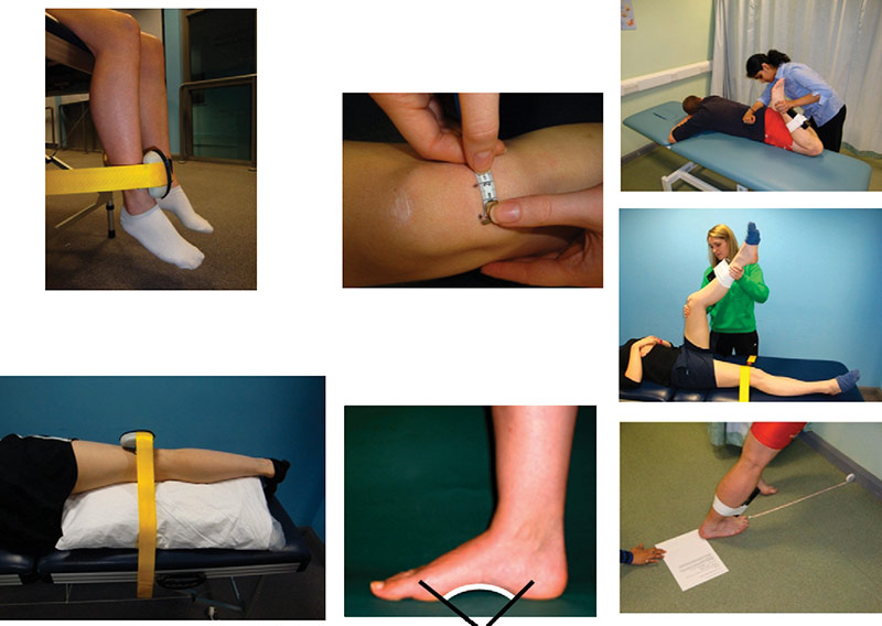
Figure 1. The seven clinical assessment tests used in the study: quadriceps strength test (top left), abductor strength test (bottom left), patella mobility test (top center), foot posture index (FPI; bottom center), and three biarticular muscle length tests (far right).
After the six potential subgroups were identified, it was time to put the assessments into clinical practice and see if subgroups would emerge. Four National Health Service (NHS) trusts participated in our study, and in total they assessed 130 people with PFP over 1.5 years. Three participants with missing data were excluded from the analysis. All participants were aged between 18 and 40 years, had patellofemoral pain in one or both legs for longer than three months, and had not yet started physiotherapy. Full details of the inclusion and exclusion criteria can be found in our two publications on this topic.11,12 Two thirds of the participants were women, which is in line with findings from others.14 On average they were aged 26 years and just on the edge of being overweight (body mass index [BMI] of 25.5). Interestingly, on average, people had PFP symptoms for 3.75 years before they saw a physiotherapist.
All participants were asked to come to one of the participating NHS trusts to be assessed. The assessment consisted of two parts. As mentioned, we wanted to look at PFP holistically; therefore, in the first part of the assessment, people were asked to complete a booklet of questionnaires to gain insight about demographic, clinical, and psychosocial aspects of PFP. This information would be used later to explore the different subgroups in more depth. The questionnaires included the Short-form McGill Pain Questionnaire, numeric pain rating scale, Leeds Assessment of Neuropathic Systems and Signs pain scale, WHO Disability Assessment Schedule, Hopkins Symptoms Checklist, movement-specific reinvestment questionnaire, self-reported indicators of cold knees, International Physical Activity Questionnaire, and modified functional index questionnaire. Prior to our study we secured approval to use the questionnaires by gaining the appropriate licenses.
The second part of the assessment consisted of the seven clinical assessment tests shown in Figure 1. A description of these clinical assessment tests is below.
A hand-held dynamometer was used to measure the weakness of the quadriceps and hip abductor muscles. The isometric quadriceps test consisted of the participant sitting on the plinth with both legs over the side. The dynamometer was fixed with a strap and placed just above the malleolus of the most affected leg. The participant was then asked to straighten this leg (knee extension). For the isometric hip abductor test the participant laid on one side with the least-affected leg on the plinth. The dynamometer was fixed with a strap and placed proximal to the knee joint of the affected leg, and the participant was asked to move the foot toward the ceiling with a straight leg (abducting the leg). The length from the hip or knee, respectively, to the point where the dynamometer was placed was recorded so the measurements could be converted to nanometers. Three attempts were recorded and the highest torque was included in the analysis.
The patellar mobility test consisted of the glide test in both the medial and lateral directions. The participant was asked to take a supine position with their legs extended and their quadriceps relaxed. First the patella was pushed to the lateral side and a mark was made on the skin where the distal point of the patella was positioned, then the patella was pushed to the medial side and the placement of the distal point of the patella was marked again with a pen. The distance between the two pen marks was then recorded, reflecting the total range of mediolateral movement. Due to this recording method, it was not possible to perform three repetitions; therefore, only one assessment was recorded per patient.

Subgrouping from clinical measures and questionnaires
Figure 2. The results of the subgroup analysis.
Foot posture was assessed once with the FPI. This test is a combination of six individual tests to assess foot posture, ranging from palpation of the talus to the congruence of the medial longitudinal arch while standing.15 This was recorded once.
The biarticular muscle length tests for the rectus femoris, hamstrings, and gastrocnemius were recorded with an inclinometer. Three attempts were recorded for each muscle and the mean length was included in the analysis. The inclinometer was fixed with a medical strap to the shin on the most affected leg. The length was measured passively. In all three muscle length tests the participant was asked if the recorded measurement was reached due to pain in or around the knee or due to muscle length. We are currently investigating if it would be better to use a goniometer for the muscle length tests, as clinicians in our first study did not like using the inclinometers despite reliable readings.
All information was collected and analyzed with respect to the published thresholds for each potential subgroup. However, no clear pattern became apparent using this approach. Then two different analytical approaches were used; hierarchical agglomerative cluster analysis and latent profile analysis. Even though these two approaches use different methods to analyze the data, the results were remarkably similar.
The subgroups
 Both approaches identified three subgroups. We have named these subgroups: “weak and tight” (39% of participants), “weak and pronated” (39%), and “strong” (22%). The weak and tight subgroup consisted of people with weak hip abductor and quadriceps muscles who also had tight lower limb muscles and hypomobility of the patella. The weak and pronated subgroup also experienced weak hip abductor and quadriceps muscles and had pronated feet (>6 on the FPI). The strong subgroup did not have weak hip abductor and quadriceps muscles but did have hypomobility of the patella. When looking from a more holistic viewpoint, it seemed that the weak and tight subgroup had the highest BMIs and were less active than the other subgroups, whereas the pronated subgroup was significantly younger than the other two. The strong subgroup had significantly higher function and quality of life than the other two subgroups, and there were proportionally more men in this subgroup (see Figure 2 for more details).

The strength measurement of the quadriceps and hip abductors and the FPI appeared to be the key clinical tests to identify the three subgroups. The mobility of the patella and the length of the quadriceps also contributed to subgroup profiles, and therefore appear to be important to consider when identifying PFP subgroups.
What’s next?
This has been the largest study to date that investigates the possibility of clinical subgroups in the PFP population. Currently, similar studies are taking place in other countries—including Turkey, Malaysia, and Thailand—to determine whether the same subgroups can be found elsewhere in patients of different ethnicities.
This is just the beginning of the identification and validation of existing subgroups in the PFP population. We assessed the participants once, but we do not yet know if there will be differences in outcome among the three subgroups after they receive treatment. Therefore, in our next step we are planning a randomized controlled trial to assess whether people with PFP will experience more improvement after receiving treatment targeting their subgroup than with conventional nontargeted therapy. Examples of these targeted treatments include the provision of a foot orthosis for people in the weak and pronated subgroup and strengthening exercises in both weak subgroups.
Jessie Janssen, PhD, is a research fellow in physiotherapy at the Allied Health Research unit at the University of Central Lancashire, UK. James Selfe, DSc, is a professor of physiotherapy at the Department of Health Professions at Manchester Metropolitan University, UK. Michael Callaghan, PhD, is a professor of clinical physiotherapy at the Department of Health Professions at Manchester Metropolitan University. Chris Sutton, PhD, is a reader in clinical trials and associate director, Lancashire Clinical Trials Unit at the University of Central Lancashire. Paola Dey, PhD, is a professor of health and social care at the Faculty of Health and Social Care at Edge Hill University, UK. Jim Richards, PhD, is a professor of biomechanics at the Allied Health Research unit at the University of Central Lancashire.
- Hill JC, Whitehurst DG, Lewis M, et al. Comparison of stratified primary care management for low back pain with current best practice (STarT Back): a randomised controlled trial. Lancet 2011;378(9802):1560-1571
- Crossley KM, Stefanik JJ, Selfe J, et al. 2016 Patellofemoral pain consensus statement from the 4th International Patellofemoral Pain Research Retreat, Manchester. Part 1: Terminology, definitions, clinical examination, natural history, patellofemoral osteoarthritis and patient-reported outcome measures. Br J Sports Med 2016;50(14):839-843.
- Selhorst M, Rice W, Degenhart T, et al. Evaluation of a treatment algorithm for patients with patellofemoral pain syndrome: a pilot study. Int J Sports Phys Ther 2015;10(2):178-188.
- Witvrouw E, Callaghan MJ, Stefanik JJ, et al. Patellofemoral pain: consensus statement from the 3rd International Patellofemoral Pain Research Retreat held in Vancouver, September 2013. Br J Sports Med 2014;48(6):411-414.
- Crossley KM. Is patellofemoral osteoarthritis a common sequela of patellofemoral pain? Br J Sports Med 2014;48(6):409-410.
- Rathleff MS, Roos EM, Olesen JL, et al. Lower mechanical pressure pain thresholds in female adolescents with patellofemoral pain syndrome. J Orthop Sports Phys Ther 2013;43(6):414-421.
- Naslund J, Naslund UB, Odenbring S, et al. Comparison of symptoms and clinical findings in subgroups of individuals with patellofemoral pain. Physiother Theory Pract 2006;22(3):105-118.
- Sheehan F, Derasari A, Fine K, et al. Q-angle and J-sign: indicative of maltracking in subgroups in patellofemoral pain. Clin Orthop Relat Res 2010;468(1):266-275.
- Harbaugh CM, Wilson NA, Sheehan FT. Correlating femoral shape with patellar kinematic in patients with patellofemoral pain. J Orthop Res 2010;28(7):865-872.
- Dierks TA, Manal KT, Hamill J, Davis I. Lower extremity kinematics in runners with patellofemoral pain during a prolonged run. Med Sci Sports Exerc 2011;43(4):693-700.
- Selfe J, Callaghan M, Witvrouw E, et al. Targeted interventions for patellofemoral pain syndrome (TIPPS): classification of clinical subgroups. BMJ Open 2013;3(9):e003795.
- Selfe J, Janssen J, Callaghan M, et al. Are there three main subgroups within the patellofemoral pain population? A detailed characterisation study of 127 patients to help develop targeted intervention (TIPPs). Br J Sports Med 2016;50(14):873-880.
- Keays SL, Mason M, Newcombe P. Individualised physiotherapy in the treatment of patellofemoral pain. Physiother Res Int 2015;20(1):22-36.
- Boling M, Padua D, Marshall S, et al. Gender differences in the incidence and prevalence of patellofemoral pain syndrome. Scandinavian J Med Sci Sports 2010;20(5):725-730.
- Redmond AC, Crosbie J, Ouvrier RA. Development and validation of a novel rating system for scoring foot posture: the Foot Posture Index. Clin Biomech 2006;21(1):89-98.


