
istockphoto.com #10411176
A delay in the activation of the peroneus longus and peroneus brevis muscles is thought to contribute to the risk of inversion ankle sprain. New research suggests that ankle taping, in addition to providing mechanical stability, also helps reduce this peroneal latency.
By Adam Knight, PhD, and Wendi Weimar, PhD
The lateral ankle sprain is one of the most common injuries in sports and was the injury reported most often in National Collegiate Athletic Association sports between 1998 and 2004.1 This injury results when the ankle is forced into excessive inversion or a combination of inversion and plantar flexion2 and commonly occurs when landing from a jump or when attempting a rapid cutting maneuver.3-6 Research has estimated that half the general population will sustain an ankle sprain during their lifetime;7 once a person sustains an ankle sprain the recurrence rate is between 70% and 80%.8,9
When the ankle is forced into inversion, the muscles and tendons that cross the foot–ankle complex on the lateral side, specifically the peroneus longus and peroneus brevis, play a key role in preventing or limiting the severity of a lateral ankle sprain.10 The muscle spindles in the peroneus longus and brevis are activated when the ankle is rapidly forced into inversion, causing a reflexive contraction of these muscles to counteract the lengthening associated with inversion.10
The time delay from initiation of the ankle inversion movement to onset of the response of peroneus longus and brevis is known as the peroneal latency, or closed loop reflex, response.11 Although there is some debate in the literature about the ability of the peroneus longus and brevis to contract quickly enough after the ankle is forced into inversion to prevent a lateral ankle sprain,12 it has been reported that the timely activation of these muscles after the ankle is forced into inversion is critical to maximizing ankle stability.13
Latency research
Until recently the preferred method in the literature to experimentally force the ankle into inversion and measure the latency of the peroneus longus and peroneus brevis has been the tilt platform.4,12,14-27 Different tilt platforms have been described in the literature, but the common methodology is a platform that has trap doors that rotate down (drop) to force the ankle into approximately 30° of inversion while a person stands quietly. However, the validity of the tilt platform for replicating the mechanics and proprioceptive cues that occur during an actual lateral ankle sprain has been called into question4,17,25 as most ankle sprains do not occur when a person is standing quietly on both legs and the floor unexpectedly falls out from under them.

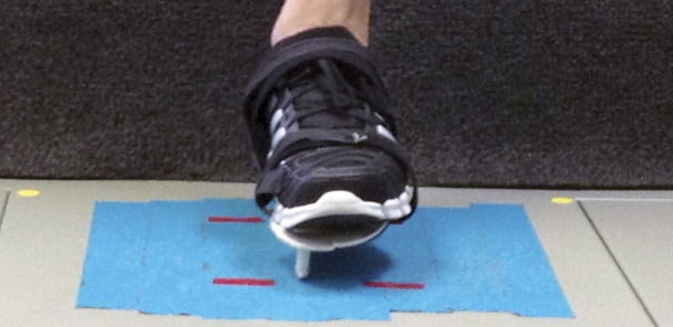
Figure 1. The authors used an outer sole with fulcrum to simulate an ankle sprain mechanism. Left: flat outer sole. Center: Outer sole with fulcrum. Right: Outer sole with fulcrum after inversion.
To create a more realistic yet safe laboratory simulation of a lateral ankle sprain, an outer sole with fulcrum was developed to force the ankle into 25° of inversion upon landing from a vertical step-down movement.28 The design of this device was taken from another study that created an outer sole with fulcrum to force the ankle into inversion and measure the effectiveness of ankle braces.29 The previous outer sole29 was made of aquaplast, contained a 27-mm high by 6-mm thick fulcrum placed 20 mm medial from the midline, and caused 24° of inversion at the subtalar joint upon landing.29 The outer sole used in the recent studies was made of orthoplast, contained a 30-mm high fulcrum placed in the same location as the design in the previous study,29 and caused 25° of inversion at the subtalar joint after landing.28,30-32 The purpose of the outer sole with fulcrum was to replicate the mechanism of a lateral ankle sprain that occurs when landing from a jump onto the foot of another player and to facilitate measurement of the latency of peroneus longus and peroneus brevis activation and the speed at which this inversion motion occurs.28,30-32
Tilt platform studies have produced differing results regarding the latency of the peroneus longus and brevis. Some studies have reported no difference in peroneal latency between healthy participants and those with ankle instability12,14,18,21,27 while others have reported increased peroneal latency among those with ankle instability.4,19,23 These studies all examined participants with some form of ankle instability. Not all people who suffer a lateral ankle sprain develop chronic ankle instability, but the recurrence rate for a lateral ankle sprain is very high.9 There is a need to investigate the effects of a single ankle sprain on the latency of the peroneus longus and brevis among participants who do not have chronic ankle instability.
Outer sole with fulcrum
Our first study investigated the effects of a previous lateral ankle sprain and previous high ankle sprain on the latency of the peroneus longus and brevis.28 Forty individuals participated in the study, including 15 with no history of any type of ankle sprain, 15 with a previous history of a lateral ankle sprain, and 10 with a previous history of a high ankle sprain. The participants performed a vertical step-down movement from a 27-cm box onto a landing surface while wearing either an outer sole with fulcrum or a flat outer sole strapped to their shoe.
The purpose of the flat outer sole was to help prevent participants from anticipating the outer sole with fulcrum by randomly strapping the flat outer sole onto his or her shoe. Surface electromyography was measured for the peroneus longus and peroneus brevis. Latency was calculated as the time (in milliseconds) from the contact of the fulcrum with the landing area (initiation of the inversion perturbation) until electromyographic (EMG) muscle activity exceeded five standard deviations (SD) from a baseline muscle activity measured 200 ms before contact.
The participants stood on top of the box and placed the testing leg behind them by extending the hip and flexing the knee. The participants were not allowed to look backwards and were instructed to look forward the entire time. Next, either the outer sole with fulcrum or flat outer sole was strapped to participants’ shoes. Participants then moved the testing foot to a position in front of, but not touching, the box. After researchers confirmed there was no EMG activity in the ankle musculature they instructed participants to step down off the box onto the testing leg.28,30-32
Results revealed no significant difference in latency between the groups for either muscle. In addition, the time to maximum inversion (TMI) was also investigated. This variable measured the time from contact of the fulcrum with the landing area until the lateral border of the outer sole made contact with the landing area, at which point the participants had completed the 25° of inversion. These results also revealed that TMI did not differ significantly between the groups.28
In a second study we investigated the effect of ankle taping on the peroneus longus latency.30 Ankle taping is commonly used to limit excessive range of motion and protect the ankle ligaments against excessive strain when forced into inversion.8 Some previous studies have reported a positive effect (a reduction in peroneus longus latency) for ankle taping,19,22 while others have reported no effect.14,24,26
For this investigation 26 participants, including 13 with no history of an ankle sprain and 13 with a previous history of a single lateral ankle sprain, completed the study protocol. The same step-down task used in our previous study28 was also used in this study. Latency was calculated as the time, in milliseconds, from the contact of the fulcrum with the landing area (initiation of the inversion perturbation) until EMG muscle activity exceeded 10 SD from a baseline muscle activity measured 200 ms before contact. Participants were tested on two separate days, 48 hours apart. On one day testing was completed without ankle taping. On the other day a certified athletic trainer taped one of each participant’s ankles using a closed basket weave technique. Participants then completed testing on the taped leg, had the tape removed, had tape applied to the opposite ankle, and completed testing on that leg. The order of tape assignment was random.
As in the previous study28 the results revealed no difference in peroneus longus latency between the two injury groups. However, there was a significant reduction in the latency of the peroneus longus across both the healthy and previously injured groups after ankle tape was applied. This reduction in latency, combined with the mechanical support offered by ankle taping, may help prevent lateral ankle sprains.30
The reduction in peroneal latency seen after application of athletic tape may be caused by an increase in cutaneous input due to the contact of the athletic tape with the skin, which could lead to an increase in the excitability of the motoneuron pool.22

We also measured the TMI and mean inversion speed (MIS) in the same study participants.31 The MIS, a measurement of the velocity of the outer sole with fulcrum, was calculated as the ratio of total angular displacement (25°) to time to maximum inversion. The participants were tested on three separate days: two days without ankle taping to measure the reliability of the TMI and MIS variables and one day with ankle taping to compare these variables across ankle support conditions and injury groups.
There were no differences between groups for TMI or MIS, findings similar to previous research measuring these variables using the tilt platform.16,27 There was also no difference in TMI and MIS between the ankle support conditions. The lack of difference in inversion speeds between the ankle support conditions seems to indicate the inversion movement during a lateral ankle sprain occurs at very high speeds, and, that over a limited range of movement of 25°, the ankle tape does not slow this speed down.

Both variables did demonstrate good reliability (for TMI, ICC = .81, for MIS, ICC = .79) and construct validity and inversion speeds measured were similar to those produced during a lateral ankle sprain that occurred during laboratory testing;33 these observations support the use of the outer sole with fulcrum methodology to replicate the mechanism of a lateral ankle sprain.31
Using the outer sole with fulcrum we also compared the latency of the peroneus longus in dominant and nondominant legs.32 It has been reported that athletes place different demands on the dominant and nondominant legs,11,34-36 and that the ankle of the dominant leg is sprained more frequently than the ankle of the nondominant leg.9,37 Fifteen healthy participants completed this study, performing 10 step downs while wearing the outer sole with fulcrum onto both the dominant and nondominant legs. Latency was calculated as the time from the contact of the fulcrum with the landing area (initiation of the inversion perturbation) until EMG muscle activity exceeded five SD and 10 SD from a baseline muscle activity measured 200 ms before contact.
Peroneus longus latency in the dominant leg was significantly greater than in the nondominant leg. This increased latency of the peroneus longus of the dominant leg may explain why the ankle of the dominant leg is reportedly sprained more frequently than the nondominant leg.32
Implications
Several conclusions can be drawn from these findings. First, utilizing the outer sole with fulcrum to force the ankle into inversion and measure the latency of the peroneus longus and peroneus brevis is an appropriate experimental tool that can safely and reliably replicate the velocities associated with an inversion sprain.31 Second, a single previous lateral ankle sprain28,30 or previous high ankle sprain does not have an adverse effect on peroneal latency.28 The effects of chronic ankle instability on peroneal latency using the fulcrum methodology are currently being investigated. Third, ankle taping can significantly reduce the latency of the peroneus longus,30 and, when combined with the mechanical support offered by ankle taping, may help prevent lateral ankle sprains. Fourth, peroneus longus latency in the dominant leg is greater than in the nondominant leg in healthy individuals, possibly increasing the risk of a lateral ankle sprain for the ankle of the dominant leg.32 Table 1 provides a summary of the findings of these studies.
Future research is needed and we are currently conducting studies to further examine the kinetics, kinematics, and EMG activity of a simulated lateral ankle sprain using the fulcrum methodology. The next step is gaining a complete understanding of the mechanics and muscular responses that occur during a lateral ankle sprain and then designing interventions that may be able to reduce the risk or number of lateral ankle sprains.
Adam Knight, PhD, is assistant professor in the Department of Kinesiology at Mississippi State University. Wendi Weimar, PhD, is an associate professor and director of the sport biomechanics laboratory in the Department of Kinesiology at Auburn University in Auburn, AL.
- Hootman JM, Dick R, Agel J. Epidemiology of collegiate injuries for 15 sports: Summary and recommendations for injury prevention. J Athl Train 2007;42(2):311-319.
- Moiler K, Hall T, Robinson K. The role of fibular tape in the prevention of ankle injury in basketball: A pilot study. J Orthop Sports Phy Ther 2006;36(9):661-668.
- McKay GD, Goldie PA, Payne WR, Oakes BW. Ankle injuries in basketball: Injury rate and risk factors. British J Sports Med 2001;35(2):103-108.
- Mitchell A, Dyson R, Hale T, Abraham C. Biomechanics of ankle instability. Part 1: Reaction time to simulated ankle sprain. Med Sci Sports Ex 2008;40(8):1515-1521.
- Ricard MD, Schulties SS, Saret JJ. Effects of high-top and low-top shoes on ankle inversion. J Athl Train 2000;35(1):38-43.
- Ricard MD, Sherwood SM, Schulties SS, Knight KL. Effects of tape and exercise on dynamic ankle inversion. J Athl Train 2000;35(1):31-37.
- Nyska M, Shabat S, Simkin A, et al. Dynamic force distribution during level walking under the feet of patients with chronic ankle instability. Br J Sports Med 2003;37(6):495-497.
- Ashton-Miller JA, Ottaviani RA, Hutchinson C, Wojtys EM. What best protects the inverted weightbearing ankle against further inversion? Evertor muscle strength compares favorably with shoe height, athletic tape, and three orthoses. Am J Sports Med 1996;24(6):800-809.
- Yeung MS, Chan KM, So CH, Yuan WY. An epidemiological survey on ankle sprains. Br J Sports Med 1994;28(2):112-116.
- Heckman DS, Reddy S, Pedowitz D, et al. Operative treatment for peroneal tendon disorders. J Bone Joint Sur Am 2008;90(2):404-418.
- Beynnon BD, Murphy DF, Alosa DM. Predictive factors for lateral ankle sprains: A literature review. J Athl Train 2002;37(4):376-380.
- Konradsen L, Voigt M, Højsgaard C. Ankle inversion injuries. The role of the dynamic defense mechanism. Am J Sports Med 1997;25(1):54-58.
- Palmieri-Smith R, Hopkins JT, Brown TN. Peroneal activation deficits in persons with functional ankle instability. Am J Sports Med 2009;37(5):982-988.
- Alt W, Lohrer H, Gollhofer A. Functional properties of adhesive ankle taping: Neuromuscular and mechanical effects before and after exercise. Foot Ankle Int 1999;20(4):238-245.
- Ebig M, Lephart SM, Burdett RG, et al. The effect of sudden inversion stress on EMG activity of the peroneal and tibialis anterior muscles in chronically unstable ankles. J Orthop Sports Phys Ther 1997;26(2):73-77.
- Eechaute C, Vaes P, Duquet W, Van Gheluwe B. Reliability and discriminative validity of sudden ankle inversion measurements in patients with chronic ankle instability. Gait Posture 2009;30(1):82-86.
- Hopkins JT, McLoda T, McCaw S. Muscle activation following sudden inversion during standing and walking. Euro J Appl Physiol 2002;99(4):371-388.
- Johnson MB, Johnson CL. Electromyographic response of peroneal muscles in surgical and nonsurgical injured ankles during sudden inversion. J Orthop Sports Phys Therapy 1993;18(3):497-501.
- Karlsson J, Andreasson GO. The effect of external ankle support in chronic lateral ankle joint instability. Am J Sports Med 1992;20(3):257-261.
- Konradsen L, Ravn JB. Prolonged peroneal reaction time in ankle instability. Int J Sports Med 1991;12(3):290-292.
- Konradsen L, Olessen S, Hansen HM. Ankle sensorimotor control and eversion strength after acute ankle inversion injuries. Am J Sports Med 1998;26(1):72-77.
- Lohrer H, Atl W, Gollhofer A. Neuromuscular properties and functional aspects of taped ankles. Am J Sports Med 1999;27(1):69-75.
- Löfvenberg R, Kärrholm J, Sundelin G, Ahlgren O. Prolonged reaction time in patients with chronic lateral instability of the ankle. Am J Sports Med 1995;23(4):414-417.
- Midgley W, Hopkins JT, Feland B, et al. The effects of external ankle support on dynamic restraint characteristics of the ankle in volleyball players. Clin J Sports Med 2007;17(5):343-348.
- Shima N, Maeda A, Hirohashi K. Delayed latency of peroneal reflex to sudden inversion with ankle taping and bracing. Int J Sports Med 2005;26(6):476-480.
- Sprigings EJ, Pelton JD, Brandell BR. An EMG analysis of the effectiveness of external ankle support during sudden ankle inversion. Can J Appl Sci 1981;6(2):72-75.
- Vaes P, Duquet W, Van Gheluwe B. Peroneal reaction times and eversion motor response in healthy and unstable ankles. J Athl Train 2002;37(4):475-480.
- Knight AC, Weimar WH. Effects of inversion perturbation after step down task on the latency of the peroneus longus and peroneus brevis. J Appl Biomech 2011;27(4):283-290.
- Ubell ML, Boylan JP, Ashton-Miller JA, Wojtys EM. The effect of ankle braces on the prevention of dynamic forced inversion. Am J Sports Med 2003;31(6):935-940.
- Knight AC, Weimar WH. Effects of ankle taping and previous injury on the latency of the peroneus longus. Sports Biomech 2012;11(1):48-56.
- Knight AC, Weimar WH. Development of a fulcrum methodology to replicate the lateral ankle sprain mechanism and measure dynamic inversion speed. Sports Biomech 2011 December 6. [Epub ahead of print.]
- Knight AC, Weimar WH. Difference in response latency of the peroneus longus between the dominant and non-dominant leg. J Sport Rehab 2011;20(3):321-332.
- Fong DTP, Hong Y, Shima Y, et al. Biomechanics of supination ankle sprain: a case report of an accidental injury event in the laboratory. Am J Sports Med 2009;37(4):822-827.
- Carey DP, Smith G, Smith DT, et al. Footedness in soccer: an analysis of France ’98. J Sports Sci 2001;19(11):855-864.
- Rahnama N, Lees A, Bambaecichi E. Comparison of muscle strength and flexibility between the preferred and non-preferred leg in English soccer players. Ergonomics 2005;48(11-14):1568-1575.
- Orchard JW. Intrinsic and extrinsic risk factors for muscle strains in Australian football. Am J Sports Med 2001;29(3):300-303.
- Ekstrand J, Gillquist J. Soccer injuries and their mechanisms: a prospective study. Med Sci Sports Exerc 1983;15(3):267-270.


