
 By Howard Kashefsky, DPM, FACFAS
Sponsored by an educational grant from Medi USA.
The terms “hallux limitus” and “hallux rigidus” refer to a degenerative process of the great toe joint that was first described by Davies-Colley1 in 1887 and termed hallux flexus. Cotterill later coined the term hallux rigidus.2 The two terms represent a progression in the spectrum of the disorder, which is characterized by gradual loss of joint range of motion (ROM) in conjunction with joint degeneration, with hallux rigidus typically defined as complete loss of motion. Athletes who experience joint loading that is both elevated and repetitive may be at risk for this problem.
Most of the current literature on hallux limitus and rigidus has focused on surgical management, especially for hallux rigidus; conservative care, however, including orthotic intervention strategies, should always be exhausted prior to surgery. In some cases a trip to the operating room seems unavoidable. Once surgery has been done, however, lower extremity clinicians may be called upon to manage the athlete postoperatively, protect a healing area, and even manage progression.
Risk factors
Hallux rigidus is the most common form of arthritis in the foot, and its incidence is increasing with an aging population.3 In the general population, hallux limitus and rigidus are more prevalent in women than in men and more likely to be bilateral than unilateral.4-6 Hallux rigidus is seen commonly in athletes, especially runners, due to repetitive stress at the great toe.7 Sports that may cause extra stress on the first metatarsophalangeal joint, including those associated with turf toe injuries such as football, basketball, and soccer (especially when played barefoot in sand), and sports that require squatting, such as the catcher’s position in baseball, set the stage for injury.8,9 In general, a soft shoe on a hard surface seems to be a risky combination for athletes where this disorder is concerned.8,9
The cause of hallux rigidus is not well understood, despite a plethora of possible causes. The most commonly reported cause is trauma—both acute trauma and repetitive microtrauma.4,10 Metatarsus primus elevates (elevation of the first ray) was first reported by Lambrinudi,11 but the question of whether this is a cause or effect of hallux rigidus remains controversial.11-15 Excessive pronation16 and internal rotation of the lower limb17 have also been linked to hallux limitus, but again, causation is difficult to determine.
Hallux biomechanics
The accepted normal ROM of the great toe joint is about 110°, and it is believed that a minimum of 65° of dorsiflexion is needed for gait.18,19 The measurement is done with the individual supine with the foot in a relaxed position and the hip and knee neutral. Passive measurement is taken with a goniometer.20 More recently, x-rays have been used to assess ROM, which may be more accurate but not always more practical.
With hallux limitus/rigidus, the proximal phalanx moves into a relative plantar position to the first metatarsal.21 The sesamoids may become retracted proximally and hypertrophied, and have visible osteophytes, joint degeneration, and ankylosis.22
Clinical manifestations
Early stages of hallux limitus injury may present as swelling, pain, edema, and limited ROM.23 Compensatory lateral foot pain and ipsilateral hip pain from external rotator muscle tightness may occur secondary to lower limb external rotation.7,24
There are several classification systems for staging.24-28 The system proposed by Coughlin and Shurnas includes grading based on actual ROM, the relative loss of ROM as a percentage of the ROM on the unaffected side, and clinical symptoms.25
In contrast, Hattrup and Johnson have developed a simple three-grade system based on radiographic findings:24
Grade 1: Mild subchondral changes with maintained joint space and minimal bony spurring.
Grade 2: Moderate subchondral changes with evidence of joint space narrowing, more bony spurring, and subchondral sclerosis or cyst.
Grade 3: Severe subchondral changes with significant narrowing, spurring, and the presence of loose fragments of cartilage or bone (see Figure 1). This is typically associated with a complete loss of range of motion.
Other classification systems for hallux limitus and rigidus have been described by Roukis et al26,27 and by Regnauld.28
Management options
In this author’s experience, surgical management of hallux rigidus in the active athlete typically involves a joint-preserving procedure rather than a fusion of the joint. A cheilectomy procedure is often done to remove bone spurs; according to Mulier, athletes in high-level sports such as judo, track and field, soccer, and skating do well with the procedure and are able to return to activity after surgery.29
However, hallux limitus can often be successfully treated conservatively. Nonsurgical options outside of shoe gear and orthoses include rest, nonsteroidal anti-inflammatory drugs, and injections of corticosteroids or sodium hylauronate.30 In a retrospective analysis of 772 patients with symptomatic hallux limitus, 55% were successfully treated—returning patients to previous activity levels with less discomfort—with conservative care alone. Of those, 84% were treated with foot orthoses, 6% with corticosteroid injections, and 10% with a change in footwear.31 Joint manipulation and other physical therapy interventions also have been reported to have benefits.32,33
Orthotic management
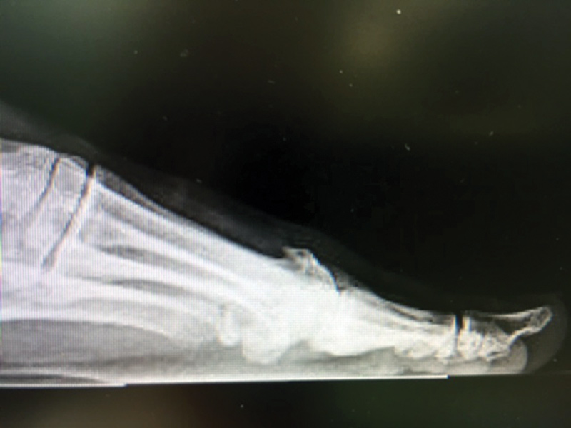
Figure 1. X-ray of an arthritic joint.
When it comes to managing this problem conservatively, orthotic devices and appropriate shoe gear play a significant role. There is, in general, a paucity of high-level evidence related to foot orthoses, and hallux limitus/rigidus is no exception; most studies involve small case series.

The goal of an orthotic device for this condition is to provide either an improvement in the postural position of the foot for mild cases or protection of the affected area for severe cases. An ideal orthotic device for the athlete will also be lightweight and low volume.
In addition, there is no clear evidence that an orthotic device for hallux limitus/rigidus needs to be a custom device. In a case series of 32 patients with first metatarsophalangeal joint pain of mechanical origin, modified prefabricated foot orthoses were associated with significantly reduced pain at 24 weeks; however, this study did not include a control group, and further analysis of a subset of patients found no association between change in pain and change in first metatarsophalangeal joint dorsiflexion.34
In this author’s opinion, an orthotic device for this patient population needs to be full contact to fit the foot appropriately, and at times this may require a custom device.
In mild cases of hallux limitus, an orthotic device may work to enhance the windlass effect, as described by Hicks.35 This effect involves shortening of the plantar fascia with dorsiflexion of the hallux and elevating the arch of the foot; conversely, it follows that a lowered arch can reduce dorsiflexion of the hallux. Hallux limitus has also been associated with pronation and plantar fasciitis, which further suggests that tension on the plantar fascia under certain conditions may influence hallux limitus.20,21 Therefore, in theory, a functional foot orthosis designed to support the mid arch in mild cases may improve symptoms.31,36
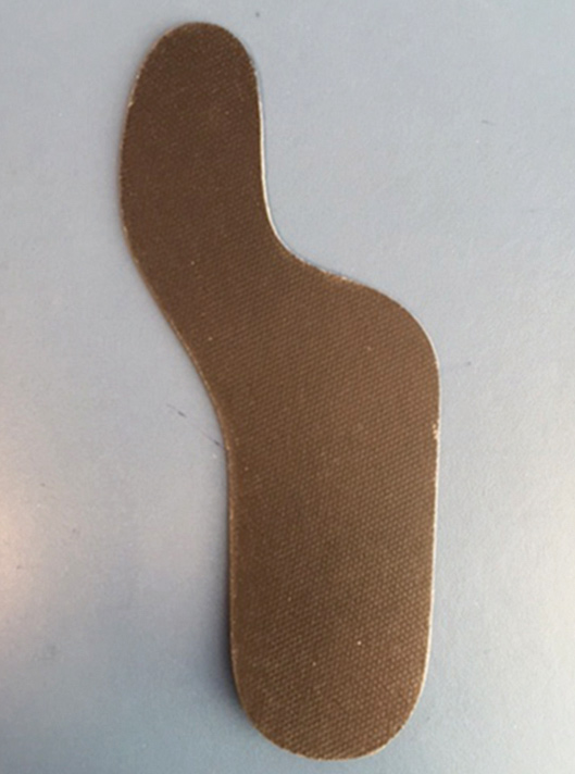
Figure 2. Rigid Morton’s extension for severe hallux rigidus
Scherer et al used this approach in fabricating custom orthoses with a 4-mm medial skive for 27 patients with functional hallux limitus, casting the feet with the first ray plantar flexed.36 They found that the mean maximum first metatarsophalangeal joint dorsiflexion during the stance phase of gait and mean maximum subhallux pressure were significantly improved when patients wore the foot orthoses compared with a no-orthosis condition.
Other options that may be helpful include a first ray cut-out, designed to allow plantar flexion of the first ray and promote further dorsiflexion of the great toe.9 Further modification to the first ray cut-out design includes a dynamic wedge inclined to 1.5 to 3 mm, elevating the second through fifth metatarsals, and a separate wedge under the distal tip of the great toe, elevating it by 1.5-3 mm, as well.37 For this modification, shoe fit in the toe box with athletic shoes may become a consideration in some circumstances.37
In contrast, in moderate to severe cases of hallux limitus/ rigidus, limiting movement of the great toe may be required to reduce symptoms. This limitation of movement could be implemented on a short-term basis to overcome acute inflammation or as a longer-term management plan. The Morton’s extension is used to extend material under the great toe to limit movement. It can be used to allow slight movement and protection when made of soft EVA (ethylene vinyl acetate). In severe cases, however, use of a much more rigid fiberglass-plate Morton’s extension may be needed (see Figure 2).9
When managing hallux limitus/rigidus, shoes and orthotic devices must work well together. The ideal shoe will have a rocker sole, be stiff enough to protect the great toe joint, and also allow for shock absorption. It should have a removable liner or allow room for an orthotic device. In the athletic population, with sport-specific shoes, this can be a challenge. Clinicians should take the time to familiarize themselves with the shoe designs and brands for each sport. Working with a team of trainers, sports medicine physicians, physical therapists, and the staff of a specialty athletic shoe store is invaluable.
Conclusion
Both hallux limitus and hallux rigidus are common injuries affecting the athletic population. Hallux limitus is a term for staging of hallux rigidus. The process is typically from trauma, either acute or repetitive microinjury. Elevation of the first ray as a contributing factor remains controversial. Depending on the severity of injury, different types of orthotic prescriptions may be appropriate. In cases of mild injury, athletes may benefit from orthotic devices that allow for protected movement. In cases of severe injury, orthoses that block movement of the first metatarsophalangeal joint may be the best option. Foot orthoses are a great tool, and in most cases may be helpful in reducing symptoms. However, more research is needed.
Howard Kashefsky, DPM, FACFAS, is the director of podiatry services at the University of North Carolina Hospitals in Chapel Hill.
- Davies-Colley M. Contraction of the metatarsophalangeal joint of the great toe. Br Med J 1887;1:728.
- Cotterill J. Stiffness of the great toe in adolescents. Br Med J 1887;1(1378):1158.
- Dellenbaugh SG, Bustillo J. Arthritides of the foot. Med Clin North Am 2014;98(2):253-265.
- Coughlin MJ, Shurnas PS. Hallux rigidus: demographics, etiology and radiographic assessment. Foot Ankle Int 2003;24(10):731-743.
- Bonney G, Macnab I. Hallux valgus and hallux rigidus: a critical survey of operative results. J Bone Joint Surg Br 1952;34(3):366-385.
- Drago JJ, Oloff L, Jacobs AM. A comprehensive review of hallux limitus. J Foot Surg 1984;23(3):213-220.
- Kunnasegaran R, Thevendran G. Hallux rigidus: nonoperative treatment and orthotics. Foot Ankle Clin 2015;20(3):401-412.
- Chou LB. Disorders of the first metatarsophalangeal joint: diagnosis of great-toe pain. Phys Sportsmed 2000;28(7):32-45.
- Tregouet P. An assessment of hallux limitus in university of basketball players compared with noncompetitive individuals. J Am Podiatr Med Assoc 2014;104(5):468-472.
- Coughlin MJ. Conditions of the forefoot. In: DeLee J, Drez D, eds. Orthopaedic Sports Medicine: Principles and Practice. Philadelphia: WB Saunders; 1994: 221-244.
- Lambrinudi P. Metatarsus primus elevatus. Proc R Soc Med 1938;31(11):1273.
- Meyer JO, Nishon LR, Weiss L, Docks G. Metatarsus primus elevatus and the etiology of hallux rigidus. J Foot Surg 1987;26(30):237-241.
- Bouaicha S, Ehrmann C, Moor BK, et al. Radiographic analysis of metatarsus primus elevatus and hallux rigidus. Foot Ankle Int 2010;31(9):807-814.
- Roukis TS. Metatarsus primus elevatus in hallux rigidus: fact or fiction? J Am Podiatr Med Assoc 2005;95(3):221-228.
- Horton GA, Park YW, Myerson MS. Role of metatarsus primus elevatus in the pathogenesis of hallux rigidus. Foot Ankle Int 1999;20(12):777-780.
- Gatt A, Mifsud T, Chockalingam N. Severity of pronation and classification of first metatarsophalangeal joint dorsiflexion increases the validity of the Hubscher Manoeuvre for the diagnosis of functional hallux limitus. Foot 2014;24(2):62-65.
- Lafuente G, Munuera PV, Dominguez G, et al. Hallux limitus and its relationship with the internal rotational pattern of the lower limb. J Am Podiatr Med Assoc 2011;101(6):467-474.
- Shereff MJ, Bejjani FJ, Kummer FJ. Kinematics of the first metatarsophalangeal joint. J Bone Joint Surg Am 1986;68(3):392-398.
- Vulcano E, Tracey JA 3rd, Myerson MS. Accurate measurement of first metatarsophalangeal range of motion in patient with hallux rigidus. Foot Ankle Int 2015 Dec 9. [Epub ahead of print]
- Aranda Y, Munuera PV. Plantar fasciitis and its relationship with hallux limitus. J Am Podiatr Med Assoc 2014;104(3):263-268.
- Flavin R, Halpin T, O’Sullivan R, et al. A finite-element analysis study of the metatarsophalangeal joint of the hallux rigidus. J Bone Joint Surg Br 2008;90(10):1334-1340.
- Munuera PV, Domınguez G, Lafuente G. Length of the sesamoids and their distance from the metatarsophalangeal joint space in feet with incipient hallux limitus. J Am Podiatr Med Assoc 2008;98(2):123-129.
- Shurnas PS. Hallux rigidus: etiology, biomechanics, and non-operative treatment. Foot Ankle Clin 2009;14(1):1-8.
- Hattrup SJ, Johnson KA. Subjective results of hallux rigidus following treatment with cheilectomy. Clin Orthop Relat Res 1998;226:182-191
- Coughlin MJ, Shurnas PS. Hallux rigidus: grading and long-term results of operative treatment. J Bone Joint Surg Am 2003;85(11):2072-2088.
- Roukis TS, Jacobs PM, Dawson DM, et al. A prospective comparison of clinical, radiographic, and intraoperative features of hallux rigidus: short-term follow-up and analysis. J Foot Ankle Surg 2002;41(3):158-165.
- Roukis TS, Jacobs PM, Dawson DM, et al. A prospective comparison of clinical, radiographic, and intraoperative features of hallux rigidus. J Foot Ankle Surg 2002;41(2):76-95.
- Regnauld B. Hallux rigidus. In Regnauld B, ed. Le Pied. Berlin, Heidelberg; Springer Verlag; 1986: 373-374.
- Mulier T, Steenwerckx A, Thienpont E, et al. Results after cheilectomy in athletes with hallux rigidus. Foot Ankle Int 1999;20(4):232-237.
- Hamid KS, Parekh SG. Clinical presentation and management of hallux rigidus. Foot Ankle Clin 2015;20(3):391-399.
- Grady JF, Axe TM, Zager EJ, Sheldon LA. A retrospective analysis of 772 patients with hallux limitus. J Am Podiatr Med Assoc 2002;92(2):102-108.
- Vallotton J, Echeverri S, Dobbelaere-Nicolas V. Functional hallux limitus or rigidus caused by a tenodesis effect at the retrotalar pulley: description of the functional stretch test and the simple hoover cord maneuver that releases this tenodesis. J Am Podiatr Med Assoc 2010;100(3):220-229.
- Shamus J, Shamus E, Gugel RN, et al. The effect of sesamoid mobilization, flexor hallucis strengthening, and gait training on reducing pain and restoring function in individuals with hallux limitus: a clinical trial. J Orthop Sports Phys Ther 2004;34(7):368-376.
- Welsh BJ, Redmond AC, Chockalingam N, Keenan AM. A case series study to explore the efficacy of foot orthoses in treating first metatarsophalangeal joint pain. J Foot Ankle Res 2010;3:17.
- Hicks JH. The mechanics of the foot. II: The plantar aponeurosis and the arch. J Anat 1954;88(1):25-30.
- Scherer PR, Sanders J, Eldredge DE, et al. Effect of functional foot orthosis on first metatarsophalangeal joint dorsiflexion in stance and gait. J Am Podiatr Med Assoc 2006;96(6):474-481.
- Rosenbloom KB. Pathology-designed custom molded foot orthoses. Clin Podiatr Med Surg 2011;28(1):171-187.


