By Brad Abicht, DPM, FACFAS
Innovative advancements are helping foot and ankle surgeons provide more precise procedures, shorten return to life activities time, and improve patient outcomes.
 Radiolucent technologies are gaining traction in foot and ankle surgeries due to their significant benefits over traditional methods. These innovations, particularly the use of carbon fiber reinforced polyetheretherketone (PEEK), are revolutionizing how surgeries are performed, offering clearer imaging and better outcomes. This article will highlight the emerging advantages of radiolucent materials in surgical procedures ranging from basic fixes to complex reconstructions.
The primary allure of radiolucent technologies in foot and ankle surgeries lies in their transparency under imaging. Unlike metal plates, which can obscure critical anatomical details, radiolucent materials such as carbon fiber reinforced PEEK allow surgeons clear visualization during and after surgical procedures. This visibility is crucial for accurate placement and adjustment during operations, and it significantly enhances the monitoring of the healing process postoperatively.
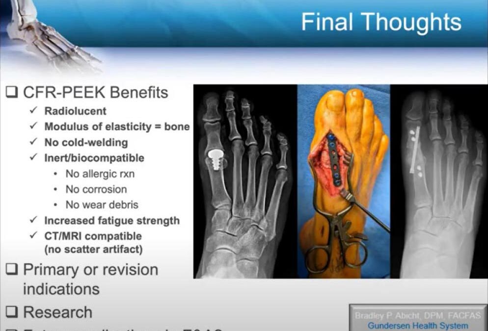
Carbon fiber reinforced PEEK is favored not only for its radiolucency but also for its biomechanical properties, which are similar to those of bone. This similarity reduces the risk of stress shielding—a common issue with metal implants where the implant takes on too much stress shielding the bone, thereby hindering optimal healing. Moreover, this material’s increased fatigue strength and compatibility with CT and MRI scans make it an excellent choice for internal fixation in orthopedics. Notably, carbon fiber materials do not cause allergic reactions, a common concern with metal-based implants, ensuring a safer postoperative experience for patients sensitive to metals.
Overview of Radiolucent Materials
Radiolucent materials are used in medical applications to allow the clear visualization of body structures during imaging procedures such as X-rays, CT scans, and MRIs. These materials are designed to be permeable to radiation, thus lacking the radio-opacity that metals possess, which typically obscures the view of the underlying biological structures. Common types of radiolucent materials include carbon fiber reinforced polymers (CFRP), such as carbon fiber reinforced PEEK. These materials are valuable in orthopedic surgeries, including those of the foot and ankle, due to their high strength, reduced artifact generation in imaging, and compatibility with the human body.
Carbon fiber reinforced PEEK offers numerous advantages in surgical applications, particularly for bone grafting and surgical treatments of the foot and ankle. One of the main benefits is its radiolucency, which significantly enhances the visibility of bones and joints during and after surgery, aiding in more accurate diagnosis, intervention, and postoperative assessment. Additionally, the material’s modulus of elasticity is similar to that of human bone, which minimizes stress shielding—a common problem with metal implants where the implant absorbs too much stress, thereby weakening the bone. Moreover, its inert nature reduces the risk of allergic reactions and inflammatory responses, which are often seen with metal-based implants. Carbon fiber reinforced PEEK also boasts high fatigue strength, which is crucial for the dynamic forces that the lower extremities endure.
Clinical Applications and Case Studies
The primary indications for using radiolucent materials in foot and ankle surgeries include scenarios where optimal visualization of bone growth and healing is necessary. This is crucial in complex reconstructive surgeries, fracture fixations, and revisions of previous surgeries. The ability to view the exact placement of bone grafts and monitor the healing process without the interference caused by metal implants presents a clear advantage in treating fractures, osteotomies, and arthrodeses. These materials have been available for other surgical uses for some time, but foot and ankle surgical indications are more recent.
Recent published studies have demonstrated the efficacy of Carbon Fiber Reinforced PEEK in various orthopedic applications. Research points to its high union rates and low complication rates compared to traditional metal plates. Systematic reviews and retrospective case reviews have supported its use, noting comparable, if not superior, outcomes in bone healing and structural stability without the complications associated with metal sensitivities and imaging artifacts.
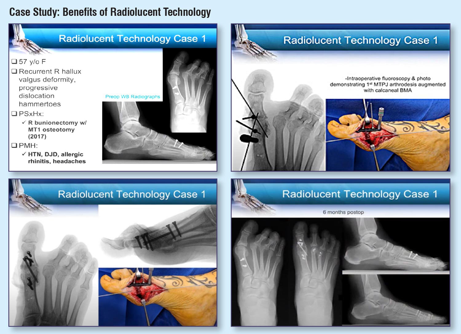
In practical applications, radiolucent materials have shown significant benefits. For instance, a case involving arthrodesis of the great toe utilized radiolucent plating, allowing unparalleled intraoperative and postoperative visualization of the joint and bone graft (see Case Study 1). This enhanced visibility aids in ensuring correct alignment and promotes more effective healing evaluations during follow-up examinations.
Another case highlighted the use of these materials in revision surgery where metallic implants had failed or caused issues. The radiolucent properties allowed for better assessment and adjustment during the procedure, which is particularly valuable in complex cases where precision is critical for successful outcomes. In revision surgeries involving bone grafting, the ability to monitor the integration of the graft visually can significantly impact the decision-making process regarding weight-bearing and rehabilitation timelines.
Advantages of Radiolucent Technologies
Improved Intraoperative Visualization
Radiolucent materials, such as carbon fiber reinforced polymer (CFRP), offer significant advantages during foot and ankle surgeries by enhancing intraoperative visualization. These technologies allow surgeons to see the bone, joint, arthrodesis, and implant directly through the radiolucent material. This superior visibility facilitates more accurate adjustments during surgery, such as optimal alignment of osteotomies and precise placement of bone grafts. Better intraoperative decisions potentially lead to improved surgical outcomes and reduced need for future corrective procedures.
Enhanced Postoperative Monitoring
After surgery, the ability to effectively monitor bone healing and implant positioning is crucial for patient recovery. Radiolucent technologies are beneficial because they do not produce the scatter artifact seen with traditional metal implants during imaging processes like X-rays, CT scans, or MRIs. As a result, clinicians can observe the healing process more clearly and make informed decisions about postoperative care, such as when to initiate weight-bearing activities or return to daily tasks. This kind of monitoring not only helps in assessing the patient’s progress but also in planning any necessary interventions at earlier stages.
Combining Radiolucent Technologies with Bone Grafting
In primary procedures involving foot and ankle surgeries, combining radiolucent technologies with bone grafting has shown to be highly effective. For instance, during arthrodesis of the great toe joint, radiolucent implants facilitate the direct visualization of the bone graft and the fusion site. This ensures that surgeons can verify the correct positioning and integration of the graft material during the initial procedure, leading to better stabilization and alignment of the joint, which are critical for successful bone healing and functional recovery.
The utility of radiolucent technologies becomes even more pronounced in revision surgeries. In complex cases where previous treatments might have failed or led to complications, the transparency of radiolucent materials allows surgeons to accurately assess and place bone grafts, ensuring proper contact and integration. For example, in the revision of a failed bunionectomy, the use of radiolucent technology enables the surgeon to precisely monitor the placement of bone grafts and assess their incorporation into the host bone over time. This can be vital for achieving successful outcomes in surgeries that aim to correct or salvage previous procedural failures.
Challenges and Considerations

While radiolucent technologies offer significant advantages in foot and ankle surgery, they also come with inherent limitations. A notable drawback is the inability of these materials to be contoured or bent during surgery. They must precisely fit the bone’s anatomy, which can be restrictive and may require additional modifications by the surgeon. Another issue arises from their radiolucency; while it enhances certain aspects of visualization, it can also complicate the precise placement of hardware as surgeons may struggle to identify the exact location of plates under X-ray or fluoroscopic guidance.
Given these limitations, addressing potential surgical complications becomes crucial. The rigidity of radiolucent materials can lead to difficulties during application, making it essential for surgical teams to have thorough preoperative planning and intraoperative precision. Additionally, the lack of ability to alter the shape of the implants on-the-fly necessitates precise bone modification, which can increase operative time and complexity. To mitigate these issues, surgeons must be highly skilled in using these materials and may benefit from advanced imaging techniques and specialized training sessions to enhance their familiarity with the characteristics and handling of radioluinecent technologies.
Future Directions and Potential Expansions
The field of radiolucent technology in orthopedic surgery is rapidly evolving, with continuous advancements aimed at overcoming current limitations. Future innovations may include the development of adjustable radioluinecent materials that can be modified during surgeries to fit individual anatomical variations precisely. Additionally, the integration of these materials with augmented reality and real-time imaging could provide surgeons with enhanced capabilities to see and navigate the surgical site more effectively. This could dramatically improve the precision of implant placement and the overall outcomes of foot and ankle surgeries.
The adoption of radio-opacities in elective foot and ankle procedures presents a unique avenue for research, particularly in the optimization of surgical outcomes and patient recovery processes. Investigating the long-term outcomes of using radiolucent materials compared to traditional metal implants can provide valuable data on the effectiveness and safety of these technologies. Furthermore, exploring patient recovery timelines, the incidence of postoperative complications, and the quality of life post-surgery could offer insights into the broader applications of radioluinecent materials in elective procedures. This research will be crucial in defining the future role of these emerging technologies in foot and ankle orthopedics.
Conclusion
The world of foot and ankle surgery stands on the brink of a transformative era thanks to the integration of radiolucent technologies and advanced bone grafting techniques. Radiolucent materials, particularly carbon fiber reinforced polymers, offer distinct advantages over traditional metal plates, including improved intraoperative visualization, enhanced postoperative monitoring, better elasticity similar to natural bone, and reduced risk of allergic reactions.
These advancements not only enable surgeons to perform procedures with greater precision but also improve patient outcomes by allowing more accurate bone healing assessments and timely interventions during the recovery phase. Additionally, the compatibility of these materials with imaging technologies like CT and MRI reduces the interference that typically complicates diagnostic processes.
As the medical community continues to embrace these innovative technologies, further research and clinical trials will undoubtedly refine their applications, making foot and ankle surgeries safer, less invasive, and more effective.
To learn more about “Emerging Radiolucent Technologies and Foot/Ankle Bone Grafting Techniques,” see Brad Abicht’s full lecture from the 38th Annual No-Nonsense Seminar, available at https://nononsense2024.lerexpo.com/.
Brad Abicht, DPM, FACFAS, is Chair of Podiatric Medicine and Surgery at Gundersen Health System in La Crosse, Wisconsin.