
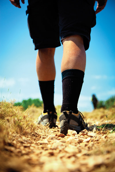
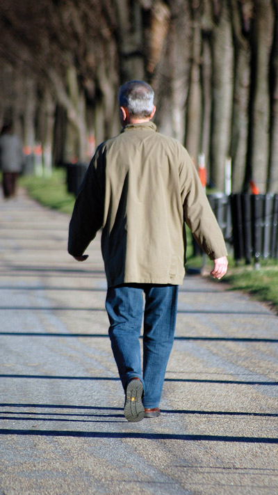
 Researchers have identified gait alterations in patients with diabetic peripheral neuropathy but also in diabetic patients with normal sensation, raising questions about the extent to which factors other than neuropathy might also be affecting gait.
By Cary Groner
Clinicians generally understand the effects of diabetic peripheral neuropathy on gait, in that patients tend to walk more slowly and with a wider stance than those without neuropathy.1 Because such gait characteristics are also found frequently in diabetes patients without sensation loss, however, researchers are raising questions about the extent to which these and other changes are associated with neuropathy per se, or are indicative of a more general diabetes-related syndrome.
It’s important, because whether they have neuropathy or not, patients with diabetes are at higher risk of gait-related problems, including foot ulcers and falling; neuropathy merely increases that risk.2,3
Research findings, as it turns out, are sometimes contradictory, but are nevertheless upending conventional thought about diabetes and gait.
“I don’t know what’s behind these gait changes, and I want to know,” said Lara Allet, PhD, a professor of physiotherapy at the University of Applied Sciences of Western Switzerland in Geneva. “I think there is something going on before peripheral neuropathy is clinically detectable, but I also think neuropathy makes the problem more severe.”
One of Allet’s theories is that the gait changes arise due to patients’ deterioration in torque development—how quickly they can activate their muscles, particularly in response to perturbations. Such capabilities have implications not only for gait but for the kinds of compensations that prevent falls.
Smita Rao, PT, PhD, an assistant professor of physical therapy at New York University in New York City, has similar questions.
“Is the gait pattern we see in diabetes driven by neuropathy?” she asked. “Is it an attempt to increase stability, reduce some of the dynamic challenges that come from walking at faster speeds or with longer stride lengths? Or is it, say, an attempt to reduce loading on the forefoot, or deal with a limited range of motion at certain joints?”
Because such factors coexist, Rao said, it’s hard to tease out the independent contributions of each. In her research, for example, she’s determined that reduction in frontal plane calcaneal mobility is an important marker for a loss of foot flexibility in diabetic patients with neuropathy.4 That flexibility loss was associated with just the kinds of gait compensations clinicians often see in patients with
diabetes.
“We found that subjects who, when tested clinically, were really limited in ankle motion actually compensated well when they walked,” Rao continued. “They limited their stride, so they walked at slower speeds, with shorter stride length. That may have been a mechanism by which they could address their ankle stiffness.”
Reduced ankle range of motion (ROM) does appear to be linked to diabetic gait characteristics, but the picture is likely more complex than that, Rao acknowledged. Researchers are beginning to grasp that complexity, however, and their new understanding
already has important implications for clinical practice.
Cautious strategies
 “In terms of joint movement, you do start to see differences even before significant neuropathy has set in,” said Neil Reeves, PhD, a senior research fellow at the Institute for Biomedical Research into Human Movement & Health at Manchester Metropolitan University in the UK.
Reeves, who is running a three-year project to investigate biomechanical and neuromuscular factors that may compromise the safety of patients with diabetes, agreed that though some aspects of diabetes-related gait are well documented, the reasons for them remain murky.
“It’s well known that these patients have a more cautious strategy,” he said. “They walk more slowly; they take shorter strides and with a lower cadence. They tend to have a wider stance, as well, which they may feel enhances stability. They may pull the hip through the stride rather than pushing off with the calf muscles. Their spatial and temporal parameters of stride also are much more variable than in people without diabetes, and that variability is probably one factor that increases their risk of falling.”
Reeves, as noted, believes such changes likely begin relatively early in the disease process and reflect its progressive nature. One way diabetes may affect gait, for example, has to do with its effect on muscle function and atrophy.
“Diabetes has a large effect on muscle function, and diabetes patients are weaker, generally, than others,” Reeves continued.5 “The disease could impact the nerves that supply the muscle, or it could impact the muscle directly by impairing the contractile machinery. Some of these gait strategies could enable patients to cope with that weakness, and that may well explain why we’re seeing changes before the onset of severe neuropathy.”
Although strength likely plays an important part, Reeves noted that even subtle sensation loss influences proprioception, which in turn may affect gait.

“I think it’s a mix of sensory and motor dysfunction,” he explained.
Echoing Smita Rao, he noted that limited ankle ROM is almost certainly part of the picture.
“That range of motion is limited because of the effects of glycation, which stiffen the muscle and tendon at the back of the leg,” he said. “That means that people can’t dorsiflex their ankles, so, when they plant their foot, the tibia won’t roll over the foot as well. As a result, they don’t progress through the stride as easily; they spend longer in a certain area, probably around the metatarsal heads, which affects the pressures placed on the foot, and in turn, the development of ulcers.”
Research
Recent research supports many of these assertions, highlights the gait changes that occur regardless of the presence of neuropathy, and seeks to explain them.
For example, a 2008 systematic review in Diabetes Metabolism Research and Reviews reported general professional consensus that diabetic patients walk slower, with greater step variability and higher plantar pressure, than healthy controls. And indeed, some of the gait abnormalities associated with diabetes were also associated with increased fall risk.6 More specifically, the studies reviewed found that diabetes was associated with adverse effects on spatiotemporal gait parameters, including velocity, stride length, gait time cycle, and single-leg support time. Other findings included greater gait variability; slower reaction times; less mobility, movement, and power in the ankles; and changes in ground reaction forces during walking. Although many of these problems were associated with peripheral neuropathy, not all were.
Investigators at Rosalind Franklin University in Chicago published a literature review in 2010 reinforcing the view that patients with diabetes often exhibit these gait patterns. But there was more to the story: such patients also showed the effects of glycosylation in the lower extremities, including decreases in skin thickness, increases in skin hardness, thickened tendons, muscle atrophy and activation delays, and limited joint ROM.7
Neuropathy has attracted its share of attention, of course. In 2011, Brazilian researchers reported that, compared with individuals without diabetes, patients with diabetic neuropathy had less ankle dorsiflexion while ascending stairs and less plantar flexion while descending. The reduced dorsiflexion compromised proper ankle and knee positions necessary for efficient lifting by the vastus lateralis; in descending, lower tibialis anterior activity may have impaired impact absorption.8 And last year, German investigators reported that intradermal injections of an anesthetic to temporarily reduce cutaneous plantar sensation to the level of neuropathy affected lower-limb kinematics and gait dynamics and contributed to modified muscle-activation patterns in healthy volunteers.9
But, as already noted, neuropathy doesn’t tell the whole story. An article from Belgian researchers this year reported that, compared with controls, older patients with diabetes walked more slowly, took shorter strides, and exhibited more gait variability—and that gait patterns did not differ between diabetic patients with and without neuropathy.10 The authors also noted that gait was affected by cognitive impairment.
Results from the Baltimore Longitudinal Study of Aging showed in 2011 that, compared with nondiabetic controls, type 2 diabetes patients without overt neuropathy spent a longer percentage of the gait cycle in the phase associated with the first occurrence of knee flexion. During fast walking, moreover, they exhibited shorter stride length and smaller hip ROM, as well as lower ankle generative mechanical work expenditure (MWE) and higher knee absorptive MWE.11
A 2012 paper in Gait & Posture tracked the electrical activity of six lower-limb muscles—the gluteus medius, rectus femoris, tibialis anterior, peroneus longus, gastrocnemius lateralis, and extensor digitorum communis—and noted an early peak of rectus femoris activity in patients with diabetes regardless of whether they had neuropathy. Furthermore, individuals without neuropathy had a midstance delay of gastroc activity, as well as delays in rectus femoris and gluteus medius activity. The authors concluded there were important muscle activity deviations in patients with diabetes that were not directly related to neuropathy.12
Some findings appear to contradict others. For example, in a 2005 article, investigators at Loma Linda University in California found type 2 diabetes patients walked more slowly, and with a wider stance, than controls. That isn’t particularly surprising. However, the authors also reported that the diabetic individuals demonstrated increased flexion/extension and lateral movement of the major joints due partly to tremor in the 8 Hz and 16 Hz bands, suggesting the increased joint movement was neurological in origin rather than due to muscle weakness or loss of foot sensation (there was no sensation loss detected in the study participants).13 That year, the same team reported that both type 1 and type 2 diabetes patients took more time, and more steps, to walk a given distance than did controls, and had at least a 50% impairment in local tissue blood flow and autonomic function.14 Intriguingly, also that year, the researchers found that gait differences in type 2 diabetes patients were reversed after six months of treatment with rosiglitazone, an insulin sensitizer, implying that gait impairment was associated with cardiovascular insufficiency to the nervous system or muscle.15
The holistic view
It’s not unusual for research in any field to raise as many issues as it answers, of course. What’s encouraging in this case is that experts are arriving at a better understanding of the breadth of the issues and redirecting their attention accordingly.
“For a long time, we focused on the neuropathic changes in diabetes, partly because they are the most apparent and you can measure them,” said Michael Mueller, PT, PhD, a professor of physical therapy at Washington University School of Medicine in St. Louis. “When you have peripheral neuropathy, you have very specific and significant impairments in sensory, motor, and autonomic nervous system areas. But what we’re seeing is that, in diabetes, you can look at just about any system and find changes. If you look for vascular changes, you’re going to find them. If you look for musculoskeletal changes, you’ll find them. Diabetes has global effects, and they affect different people in different ways.”
One suspect that has garnered a lot of attention recently is a class of compounds called advanced glycosylated end products, or AGEs, which result from elevated intracellular glucose levels and affect protein synthesis and degradation, among other things.16
“I think AGEs affect all of the musculoskeletal system, particularly the collagen in connective tissue, in muscle, in ligaments, and in joint capsules,” Mueller said.
He likened the process to one more commonly associated with holiday dinners.
“When you cure a ham, that tissue is essentially emulsified in a high concentration of glucose,” he explained. “In people, the metabolic end products of those oxidative processes are AGEs, and as a result the affected tissues become thicker, stiffer, and mechanically weaker.”
Research has elucidated this process in normal aging. Glycation-related cross-linking of intramuscular connective tissue appears to contribute to altered muscle force transmission and muscle function.16 Moreover, the longer that protein residues remain in contact with glucose, the greater the probability that AGEs will form. It doesn’t take a great leap to extrapolate the process to people with diabetes.
“It’s a normal part of aging, but people with diabetes have a much higher level of AGEs than the rest of us,” Mueller said.
Others share the view that the effect of diabetes on gait is part of a physiologically global phenomenon.
“Diabetes affects the system as a whole,” said Joseph LeMaster, MD, MPH, a professor at the University of Kansas Medical School in Kansas City. LeMaster was one of the principal investigators in the Feet First study, which showed that weight-bearing physical activity didn’t increase fall risk in patients with diabetic neuropathy—although it didn’t significantly improve balance or lower-extremity strength, either.17
“You’ve got problems with sensation and proprioception, you’ve got problems with collagen making the ankle stiffer,” he continued. “All these things together affect gait, but it’s difficult to sort out because it’s tied up with reduced balance, which is a very important part of the picture.”
Narrowing the beam
Lara Allet, the Swiss researcher mentioned earlier, has conducted extensive research to understand these issues.
In one study, published in Diabetes & Metabolism in 2009, she and her colleagues studied diabetic patients’ differences in gait speed (a performance measure) and gait variability (a fall risk) on changing terrains.18 When patients transitioned from a flat pathway to cobblestones, they slowed from ~4.5 km/hr to ~3.5 km/hr, and 36% of the decrease was associated with mean maximal isometric lower limb strength, fear of falls, and perceived vibration threshold (22%, 7.4%, and 6.4%, respectively). Moreover, variability (expressed as “coefficient of variation of the gait cycle time,” or CVGCT) rose from 2.6 to 5.1, and 11.8% of the difference was explained by mean maximal limb strength.
“We found that strength does matter, and proprioception, and fear of falling,” Allet told LER. “Now I’m trying to find out what’s going on with strength—which muscles are related to these gait problems. Is it related to strength in the ankle, knee, or hip? Is it more in the frontal plane or the sagittal plane? Is it maximum strength, or just response time?”
Allet likes to conduct her studies on uneven terrain. This is partly because she feels it accentuates gait problems and makes them easier to measure; it’s also because even though most studies are conducted on flat surfaces, in older people most falls occur on irregular ones.
In a 2009 study, she and her team assessed 45 individuals’ gait on three surfaces—tar, grass, and stones.19 Thirty subjects had type 2 diabetes (15 with neuropathy, 15 without), and there were 15 controls. There were significant gait differences between the controls and the diabetes patients, but none related to neuropathy status between the two diabetes cohorts.
In a 2012 study, Allet and her colleagues examined which frontal plane lower limb sensorimotor functions predicted gait speed and efficiency on an uneven surface in 33 older type 2 diabetes patients with neuropathy.20 They reported that frontal plane motor function at the hip and ankle (adduction and inversion, respectively) explained nearly three-fourths of the variance in gait speed, and almost half of the variance in efficiency. More specifically, maximal rate of torque development (RTD), rather than maximum strength, was most important to gait speed and efficiency. Ankle proprioceptive function did not appear to influence gait after adjusting for frontal plane motor function and confidence.
And regarding ankles, another paper, authored by Allet’s associates from the University of Michigan in Ann Arbor, used nerve conduction studies to evaluate ankle sensorimotor function relative to frontal plane postural control in patients with diabetic neuropathy compared with controls.21 They found fibular motor amplitude was associated strongly with ankle inversion/eversion proprioception and moderately associated with ankle inversion/eversion RTD, both sensorimotor capacities that influence lateral balance and recovery from perturbation during gait. The study suggested the objective nature of the measurements would offer clinicians a better way to assess proprioceptive function in such patients than methods that are common in practice today.
In the clinic
Clinical applications, of course, are paramount to practitioners and their patients, and understanding the issues is critical to developing new therapeutic approaches.
“We understand, now, that there are several reasons people have gait problems—muscle strength, proprioception, issues with metabolism,” Allet said.
She’s convinced that clinicians should be addressing gait in diabetes patients, particularly those with a history of foot ulcers.
“Do they have a gait pattern that leads to pressure at one point and not another?” she asked. “Is it because they can’t feel that they’re always stressing the same point, or is it because the gait pattern tends to overstress that point? It may be both, and if so, we shouldn’t be analyzing pressure points as isolated events, but rather together with gait issues.”
Efforts to affect gait directly have shown some success, but clinicians remain unconvinced. In a study published in the Archives of Physical Medicine & Rehabilitation, for example, a “hip walking strategy” reduced forefoot peak plantar pressures by 27%; kinematic changes included decreased plantar flexion angular velocity, hip extension ROM, and step length, as well as increased dorsiflexion and hip flexion ROM.22
Michael Mueller, one of the authors of that study, noted inherent difficulties in such approaches, however.
“We’ve suggested changing people’s walking strategies, but those changes are really hard to implement,” he said. “I think investigators will continue to try to develop sensory feedback, and get better at providing it in a more physiological or meaningful way.”
Biofeedback has shown some promise, in fact. In a 2010 study, the technique helped patients with neuropathy change their gait enough to help offload at-risk plantar surfaces. The sample was small (13 participants) and success was defined as achieving just three consecutive walking cycles of 10 steps within a target plantar pressure range.23 However, a slightly larger follow-up study, published this year, found similar effects in 21 patients that were retained at 10 days.24
More comprehensive interventions, though in their infancy, have also been promising. In one of Allet’s studies, for example, group physical therapy training including gait and balance exercises helped type 2 diabetes patients with neuropathy increase habitual walking speed on uneven terrain by .54 km/h (11.6%); cadence contributed 80% of the increase, whereas stride length constituted 20%.25 Patients in the intervention group also significantly improved their dynamic balance, performance-oriented mobility, degree of concern about falling, hip and ankle plantar flexor strength, and hip flexion mobility. Moreover, all but one of the gait and balance variables remained significantly improved at six-month follow-up. Controls, who received no treatment, demonstrated no improvement on any of the measured variables.
The future
Researchers will continue to shed light on the complex physiological processes that contribute to gait and balance problems in patients with diabetes, regardless of neuropathy status. As they do so, clinicians and their patients will doubtless benefit as this more thorough understanding leads to increasingly effective intervention strategies.
Cary Groner is a freelance writer based in the San Francisco Bay Area.
1. Khan T, Guberman R. Gait alterations associated with diabetic neuropathy. LER 2012 4(8):47-52.
2. Diabetes Public Health Resource. National Diabetes Fact Sheet, 2011. Centers for Disease Control and Prevention website. http://www.cdc.gov/diabetes/pubs/factsheet11.htm. Accessed April 19, 2013.
3. Maurer MS, Burcham J, Cheng H. Diabetes mellitus is associated with an increased risk of falls in elderly residents of a long-term care facility. J Gerontol 2005;60(9):1157-1162.
4. Rao S, Saltzman CL, Yack HJ. Relationships between segmental foot mobility and plantar loading in individuals with and without diabetes and neuropathy. Gait Posture 2010;31(2):251-255.
5. Anderson H, Nielsen S, Mogensen CE, Jakobsen J. Muscle strength in type 2 diabetes. Diabetes 2004(53):1543-1548.
6. Allet L, Armand S, Golay A, et al. Gait characteristics of diabetic patients: a systematic review. Diabetes Metab Res Rev 2008;24(3):173-191.
7. Wrobel J, Najafi B. Diabetic foot biomechanics and gait dysfunction. J Diabetes Sci Technol 2010;4(4):833-845.
8. Onodera AN, Gomes AA, Pripas D, et al. Lower limb electromyography and kinematics of neuropathic diabetic patients during real-life activities: stair negotiation. Muscle Nerve 2011;44(2):269-277.
9. Höhne A, Ali S, Stark C, Brüggemann GP. Reduced plantar cutaneous sensation modifies gait dynamics, lower-limb kinematics and muscle activity during walking. Eur J Appl Physiol 2012;112(11):3829-3838.
10. Roman de Mettelinge T, Delbaere K, Calders P, et al. The impact of peripheral neuropathy and cognitive decrements on gait in older adults with type 2 diabetes mellitus. Arch Phys Med Rehabil 2013 Feb 4 [Epub ahead of print]
11. Ko SU, Stenholm S, Chia CW, et al. Gait pattern alterations in older adults associated with type 2 diabetes in the absence of peripheral neuropathy—results from the Baltimore Longitudinal Study of Aging. Gait Posture 2011;34(4):548-552.
12. Sawacha Z, Spolaor F, Guarneri G, et al. Abnormal muscle activation during gait in diabetes patients with and without neuropathy. Gait Posture 2012;35(1):101-105.
13. Petrofsky J, Lee S, Bweir S. Gait characteristics in people with type 2 diabetes mellitus. Eur J Appl Physiol 2005;93(5-6):640-647.
14. Petrofsky J, Lee S, Macnider M, Navarro E. Autonomic, endothelial function and the analysis of gait in patients with type 1 and type 2 diabetes. Acta Diabetol 2005;42(1):7-15.
15. Petrofsky J, Lee S, Cuneo ML. Gait characteristics in patients with type 2 diabetes; improvement after administration of rosiglitazone. Med Sci Monit 2005;11(6):P143-P151.
16. Haus JM, Carrithers JA, Trappe SW, Trappe TA. Collagen, cross-linking, and advanced glycation and products in aging human skeletal muscle. J Appl Physiol 2007;103(6):2068-2076.
17. Kruse RL, LeMaster JW, Madsen RW. Fall and balance outcomes after an intervention to promote leg strength, balance, and walking in people with diabetic peripheral neuropathy: “Feet First” randomized controlled trial. Phys Ther 2010;90(11):1568-1579.
18. Allet L, Armand S, de Bie RA, et al. Clinical factors associated with gait alterations in diabetic patients. Diabet Met 2009;26(10):1003-1009.
19. Allet L, Armand S, de Bie RA, et al. Gait alterations of diabetic patients while walking on different surfaces. Gait Posture 2009;29(3):488-493.
20. Allet L, Kim H, Ashton-Miller JA, Richardson JK. Which lower limb frontal plane sensory and motor functions predicting gait speed and efficiency on uneven surfaces in older persons with diabetic neuropathy? Phys Med Rehab 2012;4(10):726-733.
21. Richardson JK, Allet L, Kim H, Ashton-Miller JA. Fibular motor conduction studies and ankle sensorimotor capacities. Muscle Nerve 2013;47(4):497-503.
22. Mueller MJ, Sinacore DR, Hoogstate S, Daly L. Hip and ankle walking strategies: effect on peak plantar pressures and implications for neuropathic ulceration. Arch Phys Med Rehabil 1994;75(11):1196-2000.
23. Pataky Z, de León Rodriguez D, Allet L, et al. Biofeedback for foot offloading in diabetic patients with peripheral neuropathy. Diabet Med 2010;27(1):61-64.
24. De León Rodriguez D, Allet L, Golay A, et al. Biofeedback can reduce foot pressure to a safe level and without causing new at-risk zones in patients with diabetes and peripheral neuropathy. Diabetes Metab Res Rev 2013;29(2):139-144.
25. Allet L, Armand S, Aminian K, et al. An exercise intervention to improve diabetic patients’ gait in a real-life environment. Gait Posture 2010;32(2):185-190.


