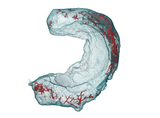
The meniscus and its network of blood vessels in a 3D rendering. The course of the blood vessels (red) is clearly visible.
The 2 menisci per knee are susceptible to wear and injury, and the meniscus is generally not a favorable candidate for a surgical procedure because blood is only supplied in certain sections. Toward this end, researchers at Empa, the Swiss Federal Laboratories for Materials Science and Technology—doctoral student Federica Orellana and principal investigator of the project, Annapaola Parrilli, PhD—are using micro- and nano-computer tomography (CT), which can even go below the micrometer limit, to create a 3D map of the cartilage. From these radiological images, the researchers create mathematical models to record and map the density, structure, biomechanical deformability, and vascular network of cartilage in space. This work could optimize treatment and enable tailored therapies in the sense of personalized medicine.
Together with clinical partners at the Istituto Ortopedico Rizzoli in Bologna, the Cantonal Hospital Winterthur, and the University of Zurich, the researchers are currently working with a large number of laboratory samples to build as meaningful a database as possible. Initial computer simulations already show the branching veins in the meniscus with promising precision. The micro-CT images convey the structural complexity of the meniscal tissue and, in the mathematical modeling, also allow further information such as the porosity or how strongly the blood vessels are tortuous.
Orellana is currently working on a 3D atlas of healthy meniscus tissue samples. In a next step, CT images of all kinds of injuries and wear and tear will be integrated into the models. The biophysicist said that the 3D map can be used for accident patients as well as for wear and tear processes in old age.


