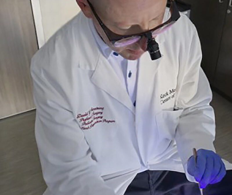
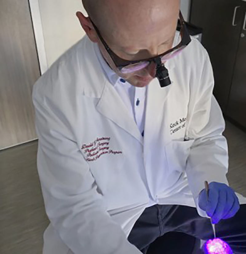
Armstrong uses AF imaging to debride a wound. Image courtesy of Armstrong.
When physicians debride a wound, such as a diabetic foot ulcer, they remove as much bacteria as possible. However, they face a key limitation—not all bacteria can be seen by the human eye, and some may be missed during the debridement. Now, Keck Medicine of University of Southern California research suggests there may be a more effective method to detect bacteria during wound debridement. Autofluorescence (AF) imaging, where a handheld device “lights up” bacteria previously invisible to the human eye, uses violet light to illuminate molecules in the cell walls of any bacteria. Different types of bacteria turn different colors, allowing physicians to immediately determine how much and which types of bacteria are in the wound.
“We’re hopeful this new technology can help surgeons improve their accuracy when pinpointing and consequently removing bacteria from wounds and therefore improve patient outcomes, particularly for those with diabetic foot wounds,” said David G. Armstrong, DPM, PhD, a podiatric surgeon and limb preservation specialist with Keck Medicine. “The early detection and removal of bacteria from a wound is vital to preventing avoidable amputations.”
According to the research, AF imaging can identify bacteria in wounds in approximately 9 in 10 patients that traditional clinical assessments miss. Additionally, with AF imaging, physicians are able to make medical decisions during the wound debridement, such as starting the patient on antibiotics or providing a special type of wound dressing, rather than waiting for lab results to initiate treatment. Another benefit to the technology is that if bacteria is caught early, the patient may avoid being prescribed antibiotics, which in wound care can be prolonged, thus avoiding possible antibiotic resistance.