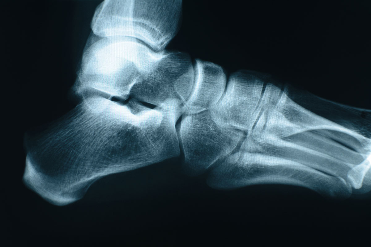
 Writing in Seminars in Musculoskeletal Radiology, 2 European experts review typical anamnestic/clinical findings, epidemiology and risk factors, imaging characteristics, and findings at typical locations of bone stress injuries in the foot and ankle that may help guide treatment strategy and patient recovery. Bone stress injuries (BSIs) are a frequent finding in athletes, particularly of the foot and ankle. A BSI is caused by recurring microtrauma to the cortical or trabecular bone exceeding the repair capacity of normal bone. The most frequent fractures at the ankle are low risk, characterized by a low risk for nonunion. These include the posteromedial tibia, the calcaneus, and the metatarsal diaphysis. High-risk stress fractures have a higher risk for nonunion and need more aggressive treatment. Examples are the medial malleolus, navicular bone, and the base of the second and fifth metatarsal bone. They note that imaging features depend on the primary involvement of cortical versus trabecular bone and that conventional radiographs may remain normal up to 2 to 3 weeks. For cortical bone, early signs of BSIs are a periosteal reaction or the “gray cortex sign,” followed by cortical thickening and fracture line depiction. In trabecular bone, a sclerotic dense line may be seen. In their opinion, magnetic resonance imaging enables early detection of BSIs and can differentiate between a stress reaction and a fracture.
Source: Jungmann PM, Schaeffeler C. Bone stress injuries at the ankle and foot. Semin Musculoskelet Radiol. 2023;27(3):283-292. doi: 10.1055/s-0043-1766098.


