
 By Cary Groner
Although footwear may in fact play a role in the development of knee osteoarthritis and its clinical management, those relationships are turning out to be much more complicated than once thought.
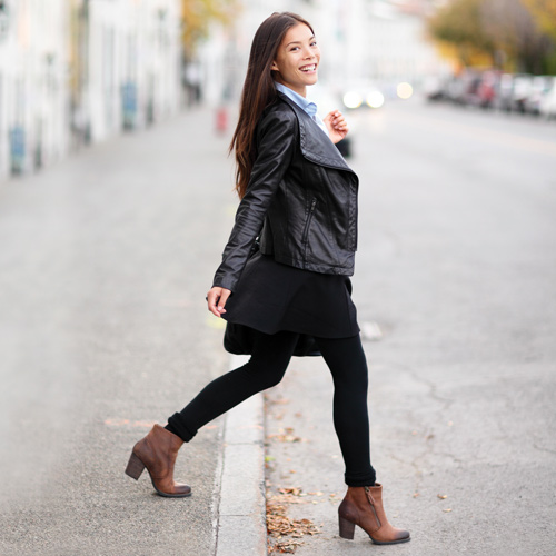
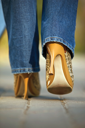
It all started with a shot across the starboard bow of high-heeled shoes. In the late 1990s, researchers began to investigate the relationship between certain types of footwear and the development and progression of knee osteoarthritis (OA). The concern was that certain shoe types—particularly those with high heels or stiff soles—could affect how loading forces passed through the knee, increasing risk for the disease. The theory gained credibility over time as more researchers expanded the work, and their findings offered hope of a noninvasive intervention that could alleviate pain and slow or stop OA’s progress.
More recently, however, others have questioned the validity of these conclusions. At the heart of the dispute lies the means by which researchers measure loading forces as they traverse the knee, and to what degree those measurements accurately reflect the cartilage damage—and resulting OA—to which those forces likely contribute. It’s a devilishly difficult thing to measure directly, and even proponents of the “shoe theory” acknowledge that their techniques represent an informed guess.
In addition, researchers and clinicians are becoming increasingly sensitive to the likelihood that interventions designed to target medial knee loading may have unintended effects elsewhere in the kinetic chain or even elsewhere in the knee joint itself.
All of this suggests that, although footwear may in fact play a role in the development of knee OA and its clinical management, those relationships are turning out to be much more complicated than once thought.

What we know
Knee OA is the primary cause of chronic disability in older people1 and particularly affects those who are overweight, female, or have muscle weakness or a history of injury.2 The disease entails a progressive degeneration of articular cartilage and tissues surrounding the joint including muscle, bone, and ligament.3 And, though OA can affect any of the knee’s three compartments, it’s most common in the medial tibiofemoral compartment.4
The biomechanical forces that appear to contribute to medial compartment OA are complicated and challenging to describe. Essentially, during gait, the weight-bearing knee aligns laterally relative to the body’s center of force, and, depending on how a person is built, this transfers force through the knee joint so that certain parts take more of the jolt than others.3 The kinetics of this action are typically described as the knee adduction moment (KAM), a measure of force and torque that represents how load is distributed across the knee’s surface. KAM is generally accepted as a valid surrogate measure of the forces on the knee,5,6 and an excessive KAM indicates torque that rotates the tibia into a varus position on the femur.3 A varus knee alignment (ie, slightly bowlegged) will experience more force through the medial compartment, and that’s where the trouble starts.

The question, again, is to what extent some shoe types increase the KAM, and whether these data can be extrapolated to predict the development or progression of knee OA.
 Not just heels
In the 1990s, Casey Kerrigan, MD, then at Harvard Medical School in Boston, became curious about why women get knee OA twice as often as men.2 Footwear seemed an obvious suspect, given that designs often differ drastically between the sexes. She and her colleagues enrolled 20 healthy women in a study and reported in the Lancet that walking in high heels (2.5-inch stilettos) altered normal ankle function, which led to compensations up the kinetic chain that increased the varus torque (another term for KAM) at the knee by 23%, versus walking barefoot.7
“Very few people at the time were looking at combining the data of the kinematic and ground reaction forces to evaluate joint torque,” said Kerrigan, who is now chair of OESH Shoes, a company she started in Charlottesville, VA. “When you put on a high heel, you’re eliminating a very important part of foot function. Normally, as you walk, you land on the outside of your foot, then roll to the inside, then go back out. There’s a transfer of forces, and high heels alter that flow, compromising the foot’s ability to neutralize those forces in the coronal plane. You can see all the things the body does to try to get that ground reaction force in line with the joint all the way up from the ankle, but the farther that force is from the knee and hip joints, the higher the torques are going to be.”
Once the principle was established, Kerrigan didn’t stop at stilettos. In a 2001 paper, also published in the Lancet, she found, surprisingly, that high-heeled shoes with a broad base were even worse than stilettos.8 In that study, of 20 healthy women wearing 2.5-inch high heels, those with heels two inches wide were associated with peak knee varus torque values that were 26% higher than in the barefoot condition, while the same measure in narrower, half-inch-base heels was 22% higher than barefoot. The results seem counterintuitive—wouldn’t the body have an easier time compensating for more stable heels of equal height?—but Kerrigan believes she knows why.
“I think it’s because stilettos make it so hard to walk that you’re taking shorter, tentative steps and not generating much torque,” she said. “With a wider heel, though, you generally feel more comfortable and you take a longer stride. This is just what I’ve observed, but it’s common sense; women wear those all day long and don’t think of them as high-heeled shoes. They think that because they’re more comfortable, they’re not as hard on your knees, but that’s not the case—any heel elevation abnormally increases the loads on the knee.”
To drive the point home, Kerrigan and her colleagues published a subsequent paper showing that moderate 1.5-inch–heeled shoes increased peak knee varus torque by up to 19% versus flat shoes.9 Then, aiming their sights ever higher (or, strictly speaking, lower), they reported that cushioned running shoes (vs barefoot)10 and even arch supports (vs running shoes without them)11 also were associated with significant increases in knee joint torque. (Consensus about this last point remains elusive; for example, in 2013 Australian researchers reported that medial arch supports had no effect on KAM or pain in patients with medial knee OA.12)
Kerrigan acknowledges that joint torque is an indirect measure of actual joint contact forces but says that, if anything, they underestimate the actual forces involved. Research supports the connection, in fact. For example, in a 2002 Japanese study, investigators followed 74 elderly patients over six years and found that those with more pain and higher KAM at baseline were more likely to experience progression of medial knee OA than the others.13 A paper from California’s Stanford University researchers that year reported that patients with knee OA had significantly higher KAM than those without OA,14 and a 2004 paper in Arthritis & Rheumatism reported that greater KAM at baseline predicted chronic knee pain three to four years later.15 A 2009 review in Knee reported that KAM increased with OA severity and was directly proportional to varus malalignment, hence potentially affecting the disease process.16 In 2012, a review of 33 articles concluded that footwear was likely to increase external KAM versus barefoot walking in patients with OA, and that lateral wedging decreased external KAM in both healthy individuals and those with OA.17 Finally, another Stanford team reported this year that maximum KAM increased with heel height in healthy women.18
Rush to judge
 While all this was going on, researchers at Rush Medical College in Chicago were coming to similar conclusions. In a 2006 paper in Arthritis & Rheumatism, for example, they assessed peak joint loads in 75 patients (59 women) with knee OA as they walked while barefoot and in their everyday shoes.19 They found that peak KAM was 11.9% lower when participants walked barefoot versus shod, and concluded that even typical street shoes (there was no mention of heel heights) could detrimentally increase loads on the lower extremity joints. Although the authors were unable to conclude decisively what factors led to this result, they speculated that sole stiffness (limiting the foot’s natural flexibility) and loss of proprioceptive input likely played roles.
The paper’s lead author, Najia Shakoor, MD, told LER that she and her colleagues came to the study roundabout; they’d been trying to reduce loads in patients with knee OA using wedged orthoses, but weren’t having a lot of success.
“We were using the wedges to help them pronate, but the orthoses also had medial arch support, so we probably weren’t accomplishing much,” she said. “Then we just had them walk barefoot in the lab, and their loads were much lower, which made us wonder what their shoes were doing. We thought we should look more closely at these supportive shoes we’d been telling them to wear.”
In 2013, Shakoor and colleagues published another paper describing the effects of specialized, flexible-sole footwear (“mobility shoes”) she and her team had developed in response to their earlier findings.20 Patients with knee OA were evaluated at baseline under three conditions: walking barefoot, in their usual shoes, and in the mobility shoes; the analyses were then repeated at six, 12, and 24 weeks. Patients wore the mobility shoes a minimum of six hours a day, six days a week, during the trial. At 24 weeks, researchers found an 18% reduction in KAM compared with baseline for both walking with mobility shoes and barefoot. Curiously, patients also experienced an 11% reduction in KAM when walking in their own shoes, which the authors speculated may have resulted from biomechanical training imparted by wearing the mobility shoes.
“There are a lot of neuromuscular reflexes that may be important when we feel our foot touch the ground,” Shakoor said. “We also think that pronation may be good for people with OA, and that because so much conventional footwear has a medial arch support, it increases knee loads. The concept of pronation as a bad thing grew largely out of podiatry, and I don’t know if it’s actually that bad; but I’m in the OA world, not the sports world.”
At baseline, the mobility shoes were associated with a KAM that was significantly higher than for walking barefoot, but those differences disappeared by the six-week time point; KAM values for the patients’ own shoes were consistently higher than for the mobility shoes for all time points, despite the reductions seen relative to baseline for both conditions.
Last November, Shakoor and several colleagues presented another paper at the annual conference of the American College of Rheumatology (ACR) in Boston, in which they reported an acute KAM reduction of 6% when patients with knee OA wore flexible shoes versus their conventional shoes. They also noted that individuals whose everyday shoes were stiffest had the greatest KAM
reductions.21
Complexities
The evidence may seem substantial, but the naysayers mentioned earlier have questioned the causal relationship between OA and factors such as KAM and footwear.
For example, in a 2013 paper, German researchers used instrumented knee implants to assess the effects of four different shoe types, versus barefoot walking, on tibiofemoral contact loads.22 With shoes, regardless of type, the researchers reported an increase of forces in the medial compartment of roughly 3% to 5%, with one exception, and questioned whether such small changes would influence OA progression.
Najia Shakoor reached a different conclusion, however.
“There were only six participants,” she said. “And, in fact, three to five percent is significant because you’re measuring one moment in time; if you think about someone with arthritis over a lifetime walking with that extra knee load on a daily basis, it becomes substantial.”
Shakoor noted, moreover, that it’s difficult to significantly affect someone’s gait in the lab because they’re habituated to walking a certain way. But, in a different shoe, over time, changes often become magnified.
“I think these studies with really small numbers of subjects, which are just looking at one point in time, don’t tell you much,” she said. “It’s really hard to say what such numbers mean in terms of a lifetime for someone with OA.”
In a 2014 article, British researchers obtained lifetime data about footwear from the Genetics of Osteoarthritis and Lifestyle (GOAL) study and reported no correlation between women’s shoes and lower limb OA.23
That study, too, had significant limitations, however. For one, individuals who reported persistent use of high- and narrow-heeled shoes in early adulthood actually were less likely to have developed knee OA than others, which strains credulity in light of other evidence. The authors noted this and suggested that such individuals may have experienced enough joint pain early in their lives to prompt a switch to more comfortable shoes, or that those who persisted in wearing high heels were less likely to have other risks for knee OA, such as high body mass index (BMI) or a high-impact occupation. Another limitation was that the paper was based on retrospective self-reported data, which—as the authors acknowledged in the paper—is often unreliable. (The authors did not respond to LER’s email requests for comment.)
“They even said in their study that they couldn’t apply a causal relationship,” noted Shakoor, who added that the authors’ speculation accurately described her personal history.
“I got joint pain in my thirties, so I modified my footwear,” she said. “Now I wear much lower heels. I can’t make sense of this [article] at all.”
Shoe stability
 At the ACR conference last fall, researchers reported on the relationship between shoe stability and knee OA risk, which they assessed using data from the Multicenter Osteoarthritis Study (MOST).24 They concluded that there was no association between walking shoe stability and two-year risk of worsening cartilage damage in the knee.
“We were trying to understand how the stability of an individual’s shoe might impact worsening medial knee cartilage damage,” said Howard Hillstrom, PhD, one of the study’s authors and director of the Leon Root Motion Analysis Laboratory at the Hospital for Special Surgery in New York. “The lack of association could be because shoes themselves aren’t necessarily the reason people’s cartilage gets worse. Another possibility is that these were qualitative assessments—a yes/no for whether the shoe was flexible or stable in a particular plane—and a quantitative measurement might have been more revealing. The other factor is that everyone doesn’t wear the same shoes, and the participants in the MOST study [mean age, about 67 years] are probably wearing sensible shoes, not four-inch stilettos or steel-toed work boots.”
Despite that study’s conclusions, Hillstrom acknowledged his interest was piqued by Shakoor’s papers showing a correlation between footwear and KAM.
“That stimulated us to see if shoes could be a problem,” he said. “Shoes affect your lower extremity biomechanics, so it doesn’t seem unreasonable that they could change your knee adduction moment, as Dr. Shakoor has reported.”
Hillstrom noted that one variable for which the study couldn’t account is similar to a limitation of the GOAL study noted above; namely, it’s nearly impossible to reliably determine what types of shoes the participants wore when they were younger, and how that footwear might have affected the onset or progression of their OA prior to their enrollment in the MOST study.
Hillstrom’s coauthor, K. Douglas Gross, MPT, DPT, ScD, an associate professor of physical therapy at the MGH Institute of Health Professions in Boston, emphasized the difficulty of drawing conclusions about the association between footwear and OA risk.
“Casey Kerrigan and others have shown that heel height is associated with measures of stress at the knee such as adduction and flexion moments,” he said. “One would want to conclude that if the heel puts added stress on an already diseased knee, that can’t be good. But I don’t think we can really relate those stress measures to OA risk.”
Gross said he doesn’t think experts yet know which characteristics of shoes are protective or damaging to the longitudinal risk of knee OA.
“We were looking particularly at the risk of the development of cartilage damage, and the worsening of cartilage damage, and we didn’t find anything,” he said. “You could easily say that we looked at the wrong shoe characteristics, because we only looked at how stable the shoe was if you bent or twisted or pinched it; but at least we can say that that measurement of stability doesn’t seem related to OA risk.”
Gross is open-minded about the potential benefits of the flexible shoes developed by Kerrigan, Shakoor, and others, but said they may not differ significantly from minimalist footwear already on the market. He pointed out that the documented limitations of minimalist shoes25 could well apply to these new shoes as well.
“Are we going to prescribe minimalist shoes for Grandma when she has to go out in the New England snow?” he asked. “What if she has diabetes? Is her podiatrist going to be happy with so little protection? What if she’s at greater risk for metatarsal fractures?”
Gross noted that potential problems might not be limited to the feet, for that matter; for example, reducing the adduction moment in the medial tibiofemoral compartment could have unintended consequences elsewhere in the knee.
“I think Howard and I can be counted among those concerned that interventions intended to treat the medial knee might be damaging other parts of the knee,” he said. “If so, you have to be very careful who you assign them to.”
Evidence-based footwear designs
Both Hillstrom and Gross noted the work of Thomas Andriacchi, PhD, and his colleagues at Stanford, who for several years have been experimenting with shoes that have dual-density midsoles (see “OA research: It’s all about the shoes,” July 2009, page 51). The slightly softer medial side allows for mild pronation when walking, in turn unloading the medial compartment of the knee. Despite their skepticism of minimalist footwear generally, Hillstrom and Gross acknowledged the promise of this approach.
“If it works for any given individual patient, it could be a great, relatively low-cost and noninvasive treatment,” Hillstrom said.
The research has been well studied and indeed seems promising. In a 2008 paper, for example, Andriacchi and his colleagues reported that, versus control (constant-stiffness) shoes, the variable-stiffness shoe was associated with a lower KAM in patients with symptoms of medial knee OA.26 In 2010, they tested the shoes in an individual with an instrumented knee replacement and reported that medial compartment joint contact force was 12.3% lower than in that individual’s personal shoe, and that the change in first peak KAM (during early stance) was significantly correlated with the change in first peak medial contact force.27 Then, in a 2011 analysis of the possible mechanisms underlying this effect, the team reported that the medial shift in the center of pressure at the foot appeared to stimulate an adaptive dynamic response during gait that reduced the frontal-plane lever arm.28 In a 2014 follow-up to the previous research, they studied knee cartilage changes over five years and found that KAM had a greater influence than knee flexion moment (KFM) on femoral cartilage change, whereas KFM more strongly influenced tibial cartilage change.29
Andriacchi’s colleague and frequent coauthor Jennifer Hledic, PhD, spoke to LER about their work. Hledic (who until her recent marriage published as JC Erhart) is a research associate of orthopedic surgery at Stanford.
“We’ve found that the variable stiffness shoes reduce the adduction moment at all tested speeds—slow, medium, and fast—but that they offer the greatest amount of reduction at fast speeds,” Hledic said.
The team hasn’t seen any deleterious effects associated with use of the dual-stiffness shoes so far, but she noted that the stiffness differential of 1.5-to-1 (between the denser and less dense parts of the sole) is subtle enough that study participants didn’t notice it when standing. The researchers have found a range of responses to the shoe, however, similar to many interventions in human study participants.
“A small percentage of people have an increase in loading, so they’re our nonresponders,” Hledic said. “The majority have reductions, but they’ve ranged from small to almost twenty-five percent.”
Hledic acknowledged that measures of KAM are surrogates for contact forces at the knee, but pointed out that the 2010 study in the instrumented artificial knee supported the validity of the approach. She was particularly encouraged by the results of the 2014 paper showing the relationship between KAM, KFM, and cartilage degeneration.
“We looked at them at baseline and five years later to see how their disease had progressed,” she said. “We segmented MRIs to get three-D representations of the cartilage, and found that baseline adduction and flexion moments were related to disease progression. At the femur, the knee adduction moment was the most significant predictor, and for tibial cartilage the flexion moment was the most significant.”
Hledic said that it’s important to pay attention to both variables, because an intervention that reduces KAM might inadvertently increase KFM, increasing overall force.
“The data from that study show that gait mechanics really can influence disease progression,” she said.
Clarity
The situation is becoming clearer in its greater complexity, in other words.
In 2013, Andriacchi published an article in Arthritis & Rheumatism that delved into the thorny nuances of such evaluations.30 Referring to another paper in the same issue, which drew a correlation between valgus alignment and lateral compartment knee OA,31 Andriacchi wrote “…that a slight valgus alignment is associated with an increased incidence of lateral compartment disease raises questions regarding the meaning of the adduction moment in the broader context of the other factors that influence this measure in the analysis of knee OA.”
Andriacchi noted that people with healthy cartilage but an elevated KAM often have thicker cartilage in the medial compartment than the lateral compartment, whereas the reverse is true in those with knee OA. The differences in the responses of healthy and arthritic cartilage to cyclic loading suggest that load is not an initial cause of OA but may instead be a factor influencing rate of disease progression. That is, in people with normal cartilage that adapts well to loading, loading may not lead to arthritis; but, in those who already have arthritis, those loads may contribute to tissue breakdown.
Andriacchi also noted that KAM doesn’t represent the actual force on the medial compartment but rather the relative medial-to-lateral distribution of force across the joint, and pointed out that muscles produce most of this force.
“As such, an increase in the adduction moment does not always mean a higher medial compartment load or a lower lateral compartment load,” he wrote. “Two subjects walking with the same peak adduction moment can have difference forces on the medial compartment…if there are different patterns of muscle contraction.”
Andriacchi concluded that, although KAM is a robust marker for medial compartment OA, a reduction in KAM alone may not reflect a change in the medial compartment load if other conditions are also changed. This may explain why some interventions that produce large reductions in knee adduction moment haven’t altered the course of OA32 or lowered the medial compartment load.33
In the clinic
All of which raises the obvious issue of what, exactly, clinicians should do for their patients with regard to footwear.
“Do we tell patients to wear supportive shoes as we used to, or do we tell them to try more minimalist shoes?” asked Shakoor. “What overall design should they look for that is mechanically advantageous for their knees if they have arthritis? You have to figure out, biomechanically, how it fits the whole person—what they need for their feet relative to what they need for their knees. If I have a diabetic patient with neuropathy, I probably won’t tell them to buy a minimalist shoe. You have to balance the comorbid conditions your patients have.”
Both Kerrigan and Shakoor have developed commercial shoes that embody the principles they’ve gleaned from their research. Kerrigan’s is flat and flexible; Shakoor’s includes sole cuts for extra flex so the foot can bend easily as it rolls forward. The Stanford team has also commercialized its variable-stiffness shoe.
But, as Shakoor noted, patient management is about more than shoes.
“It’s a good question,” said Doug Gross. “You’re a little bit on your own. I’m insistent about examining the foot before I assign any intervention, which puts my own clinical approach in contrast with the way most trials are conducted.”
Patients have to be treated as individuals, he added.
“Isn’t it reasonable to expect that somebody who has a strong tendency to hyperpronate might respond differently to a minimalist shoe than somebody with a rigid, supinated foot?” Gross asked. “There’s a lot of variability in the way people with medial knee OA respond, even in their adduction moment. Some of them may have patellofemoral involvement in addition to that of their medial tibiofemoral joint. Clinicians should look at the feet before they put anything on them, and they should look at the knee before they decide on an intervention that may be exclusively beneficial for one part of the knee but potentially damaging to other parts.”
Contrary to his recently reported research findings, however, Gross remains interested in the potential for shoes to affect disease progression.
“My secret agenda is that I think shoes matter,” he said. “I think the amount of stability and cushioning they provide, and probably heel height, all contribute to the stresses and potential for damage to the knee. As a clinician, I’ve heard over and over from patients, unprompted, ‘I can’t wear these shoes anymore, they hurt my knee.’ So it stands to reason there is a relationship.”
Cary Groner is a freelance writer in the San Francisco Bay Area.
- Guccione AA, Felson DT, Anderson JJ, et al. The effects of specific medial conditions on the functional limitations of elders in the Framingham Study. Am J Public Health 1994;84(3):351-358.
- Felson D. Epidemiology of osteoarthritis. In: Brandt K, Doherty M, Lohmander S, eds. Osteoarthritis. Oxford: Oxford University Press, 1998.
- Maly M. Knee OA: Take a load off. LER 2009;1(2):18-24.
- Ahlback S. Osteoarthritis of the knee: a radiographic investigation. Acta Radiological 1968;(Suppl 277):7-72.
- Zhao D, Banks SA, Mitchell KH, et al. Correlation between the knee adduction torque and medial contact force for a variety of gait patterns. J Orthop Res 2007;25(6):789-797.
- Hurwitz DE, Sumner DR, Andriacchi TP, Sugar DA. Dynamic knee loads during gait predict proximal tibial bone distribution. J Biomech 1998;31(5):423-430.
- Kerrigan DC, Todd MK, Riley PO. Knee osteoarthritis and high-heeled shoes. Lancet 1998;351(9113):1399-1401.
- Kerrigan DC, Lelas JL, Karvosky ME. Women’s shoes and knee osteoarthritis. Lancet 2001;357(9262):1097-1098.
- Kerrigan DC, Johansson AL, Bryant MG, et al. Moderate-heeled shoes and knee joint torques relevant to the development and progression of knee osteoarthritis. Arch Phys Med Rehabil 2005;86(5):871-875.
- Kerrigan DC, Franz JR, Keenan GS, et al. The effect of running shoes on lower extremity joint torques. PMR 2009;1(12):1058-1063.
- Franz JR, Dicharry J, Riley PO, et al. The influence of arch supports on the torques relevant to knee osteoarthritis. Med Sci Sports Exerc 2008;40(5):913-917.
- Hinman RS, Bardin L, Simic M, Bennell KL. Medial arch supports did not significantly alter the knee adduction moment in people with knee osteoarthritis. Osteoarthritis Cartilage 2013;21(1):28-34.
- Miyazaki T, Wada M, Kawahara H, et al. Dynamic load at the baseline can predict radiographic disease progression in medial compartment knee osteoarthritis. Ann Rheum Dis 2002;61(7):617-622.
- Baliunas AJ, Hurwitz DE, Ryals AB, et al. Increased knee joint loads during walking are present in subjects with knee osteoarthritis. Osteoarthritis Cartilage 2002;10(7):573-579.
- Amin S, Luepongsak N, McGibbon CA, et al. Knee adduction moment and the development of chronic knee pain in elders. Arthritis Rheum 2004;51(3):371-376.
- Foroughi N, Smith R, Vanwanseele B. The Association of external knee adduction moment with biomechanical variables and osteoarthritis: a systematic review. Knee 2009;16(5):303-309.
- Radzimski AO, Mundermann A, Sole G. Effect of footwear on the external knee adduction moment – a systematic review. Knee 2012;19(3):163-175.
- Titchenal MR, Asay JL, Favre J, et al. Effects of high heel wearer and increased weight on the knee during walking. J Orthop Res 2015;33(3):405-411.
- Shakoor N, Block J. Walking barefoot decreases loading on the lower extremity joints in knee osteoarthritis. Arthritis Rheum 2006;54(9):2923-2927.
- Shakoor N, Lidtke RH, Wimmer MA, et al. Improvement in knee loading after use of specialized footwear for knee osteoarthritis: results of a six-month pilot investigation. Arthritis Rheum 2013;65(5):1282-1289.
- Shakoor N, Lidtke RH, Ferrigno C, et al. Characteristics of conventional footwear and their association with reductions in knee loading with a flexible footwear intervention. Annual meeting of the American College of Rheumatology, Boston, November 2014.
- Kutzner I, Stephan D, Dymke J, et al. The influence of footwear on knee joint loading during walking – in vivo load measurements with instrumented knee implants. J Biomech 2013;46(4):796-800.
- McWilliams DF, Muthuri S, Muir KR, et al. Self-reported adult footwear and the risks of lower limb osteoarthritis: the GOAL case control study. BMC Musculoskelet Disord 2014;15:308.
- Gross KD, Hillstrom HJ, Niu J, et al. Relation of shoe stability to risk of knee cartilage damage: the multi-center Osteoarthritis study. Annual meeting of the American College of Rheumatology, Boston, November 2014.
- Groner C. The rise and fall of minimalist footwear. LER 2014;6(7):18-24.
- Erhart JC, Mundermann A, Elspas B, et al. A variable-stiffness shoe lowers the knee adduction moment in subjects with symptoms of medial compartment knee osteoarthritis. J Biomech 2008;41(12):2720-2725.
- Erhart JC, Dyrby CO, D’Lima DD, et al. Changes in in vivo in the loading with the variable-stiffness intervention shoe correlate with changes in the knee adduction moment. J Orthop Res 2010;28(12):1548-1553.
- Jenkyn TR, Erhart JC, Andriacchi TP. An analysis of the mechanisms for reducing the knee adduction moment during walking using a variable stiffness shoe in subjects with knee osteoarthritis. J Biomech 2011;44(7):1271-1276.
- Chehab EF, Favre J, Erhart-Hledik JC, Andriacchi TP. Baseline knee adduction and flexion moments during walking are both associated with five-year cartilage changes in patients with medial knee osteoarthritis. Osteoarthritis Cartilage 2014;22(11):1833-1839.
- Andriacchi TP. Valgus alignment and lateral compartment knee OA: A biomechanical paradox or new insight into knee OA? Arthritis Rheum 2013;65(2):310-313.
- Felson DT, Niu J, Gross KD, et al. Valgus malalignment is a risk factor for lateral knee osteoarthritis incidence and progression: findings from MOST and the osteoarthritis initiative. Arthritis Rheum 2013;65(2):355-362.
- Pham T, Maillefert JF, Hudry C, et al. Laterally elevated wedged insoles in the treatment of medial knee osteoarthritis. The two-year prospective randomized controlled study. Osteoarthritis Cartilage 2004;12(1):46-55.
- Walter JP, D’Lima DD, Colwell CW, Fregly BJ. Decreased knee adduction moment does not guarantee decreased medial contact force during gait. J Orthop Res 2010;28(10):1348-1354.


