
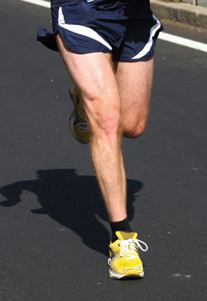
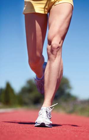
 Hip strengthening can improve short-term outcomes related to patellofemoral pain syndrome, but further research is needed to determine the underlying mechanism and whether the same approach will also prove effective in managing patellofemoral osteoarthritis.
By Michael B. Pohl, PhD
Osteoarthritis (OA) is the most common joint disease in the world.1 The National Arthritis Data Workgroup estimated in a 2008 Arthritis and Rheumatism paper that a staggering 27 million US adults were afflicted with clinical osteoarthritis2 and predicted a 40% increase in that number in the next 25 years.3 Despite its prevalence, the etiology of this disease remains unclear, and there are currently no treatments that have been proven to reverse or slow its progression.
The knee is one of the most commonly affected joints and is a major cause of pain and disability.4 The economic burden of this condition on society is significant, as it has been reported that the annual OA-related health care cost is between $6407 and $6811 per patient.5 Because knee arthroplasty and pharmacological interventions are costly to the patient and the healthcare system, it is important to develop effective conservative treatments. To achieve this, it is necessary to understand the underlying biomechanical anomalies that may contribute to disease onset and progression.
Clinicians have traditionally viewed knee OA as a disorder of the tibiofemoral joint, particularly of the medial tibiofemoral compartment. However, recent studies have discovered approximately 22% to 33% of patients with knee OA exhibit osteoarthritic changes in the patellofemoral joint6-8 and that more than a third of early knee OA sufferers have isolated involvement of the patellofemoral joint.9 Thus, patellofemoral joint osteoarthritis (PFOA) may in some instances be a precursor to tibiofemoral joint involvement.9 Additionally, compared with medial tibiofemoral OA, PFOA patients are more likely to report disability7,8 and suffer an earlier onset of chronic symptoms.7,10 Given the difference in the location of the pain and symptoms, the mechanisms leading to PFOA are likely to be different from those with primarily tibiofemoral OA. Consequently, it is also possible that patients with greater involvement of the patellofemoral joint may benefit from different rehabilitation interventions than patients with tibiofemoral OA.

Medial tibiofemoral OA has received a great deal of attention in the scientific literature in terms of its association with muscular strength deficits11 and abnormal gait patterns,12 but there is a paucity of studies exploring the relationship between strength and biomechanics in the presence of PFOA. To identify potential mechanisms, it may be useful to explore published research regarding other patellofemoral disorders.
Utting et al found that 22% of PFOA patients who were waiting to undergo a knee arthroplasty reported patellofemoral pain syndrome (PFPS) during adolescence and early adulthood.10 Moreover, given that up to 78% of PFPS patients still report chronic pain five to 20 years following rehabilitation,13-15 it seems reasonable to assume that many will go on to develop degenerative osteoarthritic changes within the patellofemoral joint. The poor long-term outcomes of PFPS, combined with the finding that many PFOA patients report preceding PFPS, suggest that the underpinning mechanical factors associated with these two conditions may be similar.
 In recent years, the hip has received attention from both the scientific and clinical communities as a potential contributor to the onset of PFPS. There is a growing body of research suggesting that PFPS patients demonstrate deficiencies in hip muscular strength. In particular, patients with PFPS have been reported to be weaker than healthy controls in both hip abduction and hip external rotation strength.16,17 A recent study has also found that individuals afflicted with PFOA were approximately 25% weaker than a healthy age- and gender-matched control group in isometric hip abduction strength18—a difference similar in magnitude to that found between PFPS patients and healthy controls.16,17
Due to the apparent decrements of hip muscular strength found in PFPS, it is not surprising that rehabilitation protocols based on hip strengthening have been gaining popularity among clinicians for the treatment of patellofemoral disorders. Significant improvements in pain and function were reported in PFPS patients who underwent an eight-week hip abductor and external rotator strengthening program.19 Studies have also illustrated that supplementing a traditional knee strengthening program with hip strengthening exercises results in greater clinical improvements than receiving the knee strengthening alone.20,21 Moreover, the former study demonstrated the additional benefits of hip strengthening may be observed 12 months post-treatment.20
From PFPS to PFOA
Assuming many individuals with chronic PFPS will go on to develop osteoarthritic changes at the patellofemoral joint,10 it seems necessary to consider studies of interventions for PFPS when developing interventions for patients with PFOA. The primary aims of the management of knee OA are to educate the patient, control pain, improve function, and slow disease progression.22 In the advanced stages of this disease, surgical interventions such as knee arthroplasty are commonly necessary, although arthroscopic surgery for knee OA has recently been shown to provide no additional benefit to physical and medical therapy.23 In the earlier stages, nonoperative conservative treatment may be more desirable. Both the European League Against Rheumatism (EULAR) and Osteoarthritis Research Society International (OARSI) recommendations propose exercise as a first-line therapy for the management of knee OA.22,24
Traditionally, exercise therapy for knee OA has generally focused on patients with predominant tibiofemoral OA or has not attempted to target disease subgroups based on the affected compartment.25,26 Moreover, exercise therapy has primarily focused on strengthening the quadriceps. To the author’s knowledge, only one study has investigated the efficacy of physical therapy in patients with predominant patellofemoral joint involvement.27 However, this study used traditional quadriceps exercises despite the shift toward hip-based strengthening in treating patellofemoral disorders. A recent pilot study has shown that a 17% increase in hip abduction strength following a six-week strengthening program in patients with PFOA resulted in significant improvements in both pain and knee-related quality of life.28 Although these results are promising, the mechanical explanation for how hip muscular weakness is related to the pain and symptoms of patellofemoral disorders is not fully understood.
It has been proposed that alterations in hip biomechanics secondary to muscular weakness may cause patellar maltracking and alter the normal loading pattern at the patellofemoral joint.29 The hip abductor and external rotator muscle groups act to oppose the motions of hip adduction and internal rotation, respectively. Excessive hip adduction and internal rotation have been shown to be present during gait in patients with PFPS.30-32 Excessive internal rotation of the femur may result in lateral patellar tilt, a phenomenon that has been demonstrated in vivo.33 Greater hip adduction may place more tension on the iliotibial band, which can cause the patella to tilt laterally and also translate laterally.34 Research has demonstrated greater magnitudes of lateral patellar tilt and translation in PFPS patients than controls during a squatting movement performed in a magnetic resonance imaging scanner.35 Further, cadaveric studies have shown that both lateral patellar tilt and translation can result in abnormal loads on the patellofemoral joint,36,37 which may lead to the discomfort experienced by PFPS patients. Therefore, strengthening the muscle groups that control the movements of hip adduction and internal rotation may be effective in the treatment of PFPS/PFOA because this helps correct patellar maltracking.
While numerous studies have investigated these abnormal gait biomechanics in patients with PFPS, the literature on gait biomechanics associated with PFOA is scarce. One recent study found no differences in either hip adduction or hip external rotation peak angles during walking in patients with mild to moderate PFOA compared with healthy controls.18 This contradicts some of the PFPS research. As discussed earlier, the PFOA group in the aforementioned study displayed significantly less hip abduction strength (25% less) than controls, suggesting the hip abductor weakness in the PFOA group had no effect on gait biomechanics during walking.18 However, it cannot be firmly concluded from these results that hip abductor weakness in general will not lead to alterations in hip biomechanics.
Walking requires significantly less hip abductor activation than the maximal isometric contraction that is typically used to assess strength. Indeed, researchers found hip adduction angle remained unchanged in healthy individuals following an experimentally induced 24% reduction in maximal hip abduction strength.38 Therefore, it is possible the reduction in isometric hip abduction strength in PFOA subjects was not sufficient to cause an increase in hip adduction during walking. Strength deficiencies may have biomechanical implications only during more demanding movement tasks such as running and jumping, which place greater demands on the musculature.
To complicate matters, Earl and colleagues39 recently found that hip adduction and external rotation during running remained unchanged following an eight-week hip and core strengthening program, despite significant improvements in symptoms and both hip abductor and external rotator muscle strength. This raises the question of how hip strengthening can improve the symptoms of PFPS without causing any measurable changes in hip biomechanics. A potential explanation is that subtle changes in gait biomechanics following an intervention cannot always be measured using conventional 3D motion analysis techniques. It is nearly impossible for even a highly trained researcher to place anatomical markers in exactly the same location during any two separate testing sessions.40 Such errors in marker placement can affect the subsequent joint angles measured during gait and the calculation of any kinematic changes.
Secondly, and more importantly, the markers are placed externally on the skin and do not necessarily represent the underlying bony motion.41 In particular, markers placed on the thigh may not accurately depict the movement of the femur. Moreover, it is currently not possible to track the motion between the patella and femur joint using motion analysis. Advancements in imaging technology, such as magnetic resonance imaging and fluoroscopy, offer promising methods of assessing patellofemoral joint motion and how it may be altered following various interventions.42 The identification of interventions that effectively correct abnormal patellofemoral joint mechanics in a PFPS population may prove valuable in terms of preventing the development of osteoarthritis at the joint.
Multiple factors
While this article has been written with a focus on the possible association between hip strength and biomechanics with patellofemoral pain, it is important to acknowledge that this condition is multifactorial in nature. Clinicians and scientists interested in PFPS gathered in 2008 in Baltimore and in 2010 in Ghent, Belgium, at the first and second International Patellofemoral Research Retreats to present new research and review existing literature regarding patellofemoral pain.43-44 (A third retreat was held in September 2013 in Vancouver, Canada; the proceedings have not yet been published.)
The groups produced consensus statements that categorized the causes of PFPS into three broad mechanistic subgroups: local factors (e.g., anatomy of the patellofemoral joint, quadriceps weakness, impaired vastus medialis obliquus function); distal factors (e.g., rearfoot eversion, tibial internal rotation); and the proximal factors discussed in this article. The majority of research to date consists of studies that compare a sample of PFPS patients to a control group and identify factors that are different between the two groups. However, any given sample may be composed of individuals with different proximal, local, or distal factors contributing to their injury, and this likely contributes to the often conflicting results found among studies.

To truly uncover the mechanisms behind patellofemoral pain, perhaps it is time to shift the approach to conducting research on the topic. Given that each subgroup of patients will have varying factors contributing to their pain, it is logical that different subgroups will require different interventions to resolve symptoms. For example, an individual who is experiencing PFPS due to hip abductor weakness would be likely to respond to a proximal strengthening program. By contrast, the odds of an individual with a local problem, such as patella alta, responding to the proximal strengthening program are much lower. It may be possible to tease out the separate subgroups by first applying a targeted intervention, and then categorizing patients based on their response to the treatment.
In summary, PFOA is a debilitating condition that may be preceded by PFPS earlier in life. The poor long-term outcomes of PFPS following rehabilitation highlight the need to develop more effective treatments to prevent degenerative osteoarthritic changes occurring at the patellofemoral joint. Hip strengthening has proven efficacious in improving short-term outcomes related to PFPS, but further work is needed to determine whether it will also prove effective in managing PFOA. The exact mechanisms by which hip strengthening reduces the symptoms associated with patellofemoral disorders are still unclear. Future research using cutting-edge imaging techniques is needed to provide insight into the biomechanics of the patellofemoral joint in PFPS and PFOA and how maltracking may be altered using various interventions.
Michael B. Pohl, PhD, is an assistant professor in the Department of Kinesiology and Health Promotion at the University of Kentucky in Lexington.
- Report of the joint disease consensus group: The bone and joint decade 2000-2010. Osteoarthritis Cartilage 1999;7(3):251-254.
- Lawrence RC, Felson DT, Helmick CG, et al. Estimates of the prevalence of arthritis and other rheumatic conditions in the United States: Part II. Arthritis Rheum 2008;58(1):26-35.
- Helmick CG, Felson DT, Lawrence RC, et al. Estimates of the prevalence of arthritis and other rheumatic conditions in the United States: Part I. Arthritis Rheum 2008;58(1):15-25.
- Oliveria SA, Felson DT, Reed JI, et al. Incidence of symptomatic hand, hip, and knee osteoarthritis among patients in a health maintenance organization. Arthritis Rheum 1995;38(8):1134-1141.
- Le TK, Montejano LB, Cao Z, et al. Healthcare costs associated with osteoarthritis in US patients. Pain Pract 2012;12(8):633-640.
- Davies AP, Vince AS, Shepstone L, et al. The radiologic prevalence of patellofemoral osteoarthritis. Clin Orthop Relat Res 2002;(402):206-212.
- Duncan R, Peat G, Thomas E, et al. How do pain and function vary with compartmental distribution and severity of radiographic knee osteoarthritis? Rheumatology 2008;47(11):1704-1707.
- McAlindon TE, Snow S, Cooper C, Dieppe PA. Radiographic patterns of osteoarthritis of the knee joint in the community: the importance of the patellofemoral joint. Ann Rheum Dis 2002;51(7):844-849.
- Kumm J, Tamm A, Lintrop M, Tamm A. The prevalence and progression of radiographic knee osteoarthritis over 6 years in a population-based cohort of middle-aged subjects. Rheumatol Int 2012;32(11):3345-3550.
- Utting MR, Davies G, Newman JH. Is anterior knee pain a predisposing factor to patellofemoral osteoarthritis? Knee 2005;12(5):362-365.
- Hurley MV. The role of muscle weakness in the pathogenesis of osteoarthritis. Rheum Dis Clin North Am 1999;25(2):283-298.
- Andriacchi TP, Mündermann A. The role of ambulatory mechanics in the initiation and progression of knee osteoarthritis. Curr Opin Rheumatol 2006;18(5):514-518.
- Nimon G, Murray D, Sandow M, Goodfellow J. Natural history of anterior knee pain: A 14- to 20-year follow-up of nonoperative management. J Pediatr Orthop 1998;18(1):118-122.
- Stathopulu E, Baildam E. Anterior knee pain: a long-term follow-up. Rheumatology 2003;42(2):380-382.
- Blond L, Hansen L. Patellofemoral pain syndrome in athletes: a 5.7-year retrospective follow-up study of 250 athletes. Acta Orthop Belg 1998;64(4):393-400.
- Ireland ML, Willson JD, Ballantyne BT, Davis IM. Hip strength in females with and without patellofemoral pain. J Orthop Sports Phys Ther 2003;33(11):671-676.
- Willson JD, Davis IS. Lower extremity strength and mechanics during jumping in women with patellofemoral pain. J Sport Rehabil 2009;18(1):76-90.
- Pohl MB, Patel C, Wiley JP, Ferber R. Gait biomechanics and hip muscular strength in patients with patellofemoral osteoarthritis. Gait Posture 2013;37(3):440-444.
- Khayambashi K, Mohammadkhani Z, Ghaznavi K, et al. The effects of isolated hip abductor and external rotator muscle strengthening on pain, health status, and hip strength in females with patellofemoral pain: a randomized controlled trial. J Orthop Sports Phys Ther 2012;42(1):22-29.
- Fukuda TY, Melo WP, Zaffalon BM, et al. Hip posterolateral musculature strengthening in sedentary women with patellofemoral pain syndrome: a randomized controlled clinical trial with 1-year follow-up. J Orthop Sports Phys Ther 2012;42(10):823-830.
- Nakagawa TH, Muniz TB, de Marche Baldon R, et al. The effect of additional strengthening of hip abductor and lateral rotator muscles in patellofemoral pain syndrome: a randomized controlled pilot study. Clin Rehabil 2008;22(12):1051-1060.
- Zhang W, Moskowitz RW, Nuki MB. OARSI recommendations for the management of hip and knee osteoarthritis, Part II: OARSI evidence-based, expert consensus guidelines. Osteoarthritis Cartilage 2008;16(2):137-162.
- Kirkley A, Birmingham TB, Litchfield RB, et al. A randomized trial of arthroscopic surgery for osteoarthritis of the knee. N Engl J Med 2008;359(11):1097-1107.
- Jordan KM, Arden NK, Doherty M, et al. EULAR Recommendations 2003: an evidence based approach to the management of knee osteoarthritis: Report of a Task Force of the Standing Committee for International Clinical Studies Including Therapeutic Trials (ESCISIT). Ann Rheum Dis 2003;62(12):1145-1155.
- Bennell KL, Hinman RS, Metcalf BR, et al. Efficacy of physiotherapy management of knee joint osteoarthritis: a randomised, double blind, placebo controlled trial. Ann Rheum Dis 2005;64(6):906-912.
- Fransen M, Crosbie J, Edmonds J. Physical therapy is effective for patients with osteoarthritis of the knee: a randomized controlled trial. J Rheumatol 2001;28(1):156-164.
- Quilty B, Tucker M, Campbell R, Dieppe P. Physiotherapy, including quadriceps exercises and patellar taping, for knee osteoarthritis with predominant patello-femoral joint involvement: randomized controlled trial. J Rheumatol 2003;30(6):1311-1317.
- Pohl MB, Wiley JP, Patel C, Ferber R. The effect of a hip strengthening program on muscle strength, gait biomechanics, pain and function in patients with patellofemoral osteoarthritis. Presented at the American College of Sports Medicine Annual Meeting, Indianapolis, IN, June 2013.
- Powers CM. The influence of altered lower extremity kinematics on patellofemoral joint dysfunction: a theoretical perspective. J Orthop Sports Phys Ther 2003;33(11):639-646.
- Noehren B, Pohl MB, Sanchez Z, et al. Proximal and distal kinematics in female runners with patellofemoral pain. Clin Biomech 2012;27(4):366-371.
- Souza RB, Powers CM. Differences in hip kinematics, muscle strength, and muscle activation between subjects with and without patellofemoral pain. J Orthop Sports Phys Ther 2009;39(1):12-19.
- Willson JD, Davis IS. Lower extremity mechanics of females with and without patellofemoral pain across activities with progressively greater task demands. Clin Biomech 2008;23(2):203-211.
- Souza RB, Draper CE, Fredericson M, Powers CM. Femur rotation and patellofemoral joint kinematics: a weight-bearing magnetic resonance imaging analysis. J Orthop Sports Phys Ther 2010;40(5):277-285.
- Merican AM, Amis AA. Iliotibial band tension affects patellofemoral and tibiofemoral kinematics. J Biomech 2009;42(10):1539-1546.
- Draper CE, Besier TF, Santos JM, et al. Using real-time MRI to quantify altered joint kinematics in subjects with patellofemoral pain and to evaluate the effects of a patellar brace or sleeve on joint motion. J Orthop Res 2009;27(5):571-577.
- Huberti HH, Hayes WC. Patellofemoral contact pressures. The influence of q-angle and tendofemoral contact. J Bone Joint Surg Am 1984;66(5):715-724.
- Lee TQ, Anzel SH, Bennett KA, et al. The influence of fixed rotational deformities of the femur on the patellofemoral contact pressures in human cadaver knees. Clin Orthop Relat Res 1994;(302):69-74.
- Pohl MB, Kendall K, Wiley P, et al. The role of experimentally induced hip abductor muscle strength deficits on frontal plane biomechanics during gait. Presented at 35th Annual Meeting of the American Society of Biomechanics, Long Beach, CA, August 2011.
- Earl JE, Hoch AZ. A proximal strengthening program improves pain, function, and biomechanics in women with patellofemoral pain syndrome. Am J Sports Med 2011;39(1):154-163.
- Pohl MB, Lloyd CH, Ferber R. Can the reliability of three-dimensional running kinematics be improved using functional joint methodology? Gait Posture 2010;32(4):559-563.
- Reinschmidt C, van den Bogert AJ, Nigg BM, et al. Effect of skin movement on the analysis of skeletal knee joint motion during running. J Biomech 1997;30(7):729-732.
- Nha KW, Papannagari R, Gill TJ, et al. In vivo patellar tracking: clinical motions and patellofemoral indices. J Orthop Res 2008;26(8):1067-1074.
- Davis IS, Powers CM. Patellofemoral pain syndrome: proximal, distal, and local factors, an international retreat, April 30-May 2, 2009, Fells Point, Baltimore, MD. J Orthop Sports Phys Ther 2010;40(3):A1-A16.
- Powers CM, Bolgla LA, Callaghan MJ, et al. Patellofemoral pain: proximal, distal, and local factors. 2nd International Research Retreat. J Orthop Sports Phys Ther 2012;42(6):A1-A54.


