If custom foot orthoses are a significant part of your practice, digital scanning technology can save time and money without compromising clinical quality – as long as the scanner provides the clinical information you need.
 By Larry Huppin, DPM
When capturing an image of the foot for production of custom foot orthoses, the majority of practitioners take a negative cast just as it has been done for the past 50 years – using plaster strips to form a non-weightbearing negative cast of the foot. Some have moved on to using polyester socks to capture the foot. Others use foam boxes. Currently a very small percentage are using digital technology to capture an image of the foot. However, there are several technologies that allow Digital Imaging Services, the most promising of which is optical scanning.
A 2007 La Trobe University cost-benefit analysis, comparing optical scanning to plaster casting, showed an overwhelming benefit to the use of optical scanners.1 Casting both feet–including preparation, casting, prescription writing and clean-up—took approximately 11 minutes with plaster vs. two minutes for an optical scanner. Cost comparisons also weighed heavily on the side of the scanner. Assuming that a practitioner’s time is worth $100 to $150(AUD) per hour, the cost per patient for a plaster cast was $27.94 to $49.60(AUD) versus $3.30 to $10(AUD) for the optical scan. These costs reflect casting time and materials, but do not include the capital cost of the optical scanner.
Given the great cost and efficiency benefits, the number of digital imagers being used is likely to grow rapidly over the next few years. I predict that that within five years, most practitioners will capture the image digitally.
If we are going to gravitate toward the use of digital imagers, we must become educated consumers and understand the advantages, disadvantages, benefits and limitations of these new technologies. Practitioners considering the purchase of such technology must ask themselves several questions. What do I need to capture to make a clinically effective foot orthosis? Will a particular digital imager capture this information? Can optimum outcomes be achieved using this technology? What are the advantages and disadvantages of the technology?
Casting criteria
The first step in evaluating digital imagers is to take a step back and look at the foot position in which the image must be captured to achieve optimum clinical outcomes. McPoil et al looked at this question in 1989 and found that foam box semi-weightbearing casting resulted in a forefoot-to-hindfoot angle that was significantly smaller than the angle measured using either of two nonweightbearing methods.2 They suggested that the difference is due to the inability of the midtarsal joint to lock using the semi-weightbearing method.
Davis et al did a similar study in 2002 where they compared methods for taking a negative cast of the foot.3 Non-weightbearing plaster was compared to semi-weightbearing foam. Their conclusions were that NWB casting had the highest level of agreement with the clinically measured forefoot-to-rearfoot relationship while SWB foam casting had the lowest level of agreement. The conclusion of the study was to recommend plaster casting as the most reliable and valid method in situations where the forefoot-to-rearfoot relationship is of importance.
These studies bring us to the first rule of negative casting:
Casting Rule #1: Non-weightbearing casting is the best method to ensure that the first ray will be plantarflexed.
If semi-weight bearing casting methods risk putting the foot in a position of increased forefoot varus, how will this affect orthoses? Roukis et al found that prevention of first ray plantarflexion resulted in decreased first MPJ dorsiflexion (hallux limitus).4 A subsequent study by Scherer et al looked at the effect in stance of a polypropylene orthosis made from a NWB negative cast taken with the first ray plantarflexed on patients with a functional hallux limitus. Results of this study were that mean dorsiflexion at the first MPJ increased 90%.5
In a 2000 study, Harradine found that increasing eversion of the heel decreased available dorsiflexion at the first MPJ.6 According to Root et al, “ground reaction against the first metatarsal head will force the first ray into a dorsiflexed position when the foot is everted by pronation”,7 and based on the study by Roukis et al it can be hypothesized that the decrease in hallux dorsiflexion is secondary to the dorsiflexion of the first ray.4
These studies indicate that first ray dorsiflexion leads to decreased hallux dorsiflexion, resulting in functional hallux limitus. Casting the foot with the first ray dorsiflexed will result in an orthotic device that holds the first ray dorsiflexed. This may contribute to increased first MPJ symptoms as a result of joint jamming.
So, the second rule of casting is:
Casting Rule #2: Orthoses made from casts with a dorsiflexed first ray are more likely to lead to jamming of the first MPJ. Those made with a plantarflexed first ray enhance first MPJ dorsiflexion.
Casting with a plantarflexed first ray may have other benefits. A cadaveric study by Kogler in 1999 found that valgus forefoot wedging decreased strain on the plantar fascia under experimental weightbearing conditions, while varus wedging increased strain.8 This study showed unequivocally that the most effective way to decrease strain on the plantar fascia is to evert the forefoot.
To provide the force needed to evert the forefoot, a valgus wedge can be added to the orthosis, but valgus should also be captured within the orthotic plate itself. To do so, as much valgus as possible should be captured during the casting process. To accomplish this, the subtalar joint should be held in neutral, the midtarsal joint maximally pronated and the first ray should be plantarflexed during casting.
Casting Rule #3: To produce an orthosis that decreases strain on the plantar fascia, cast out any soft tissue varus to enhance forefoot valgus in the negative cast.
By definition, a functional orthosis is one in which the forefoot is balanced relative to the perpendicular axis of the rearfoot in the frontal plane when the foot is held non-weightbearing in subtalar neutral with the midtarsal joint locked. Without this balancing, the orthosis is providing only a simple arch support. Kogler’s study8 demonstrated effectively the importance of frontal plane valgus wedging of the forefoot in order to decrease strain on the plantar fascia. Valgus wedging is built into a functional foot orthosis by capturing a valgus forefoot position in the negative cast and then balancing (supporting) the valgus position of the forefoot. If the forefoot valgus is not supported (balanced to the rearfoot) then the orthosis will not be optimized to decrease strain on the plantar fascia.
As far back as the late 1950s, Manter, whose work influenced Root, suggested that supination of the midtarsal joint leads to destabilization of the rearfoot.9 This would suggest that a functional orthosis that stabilizes the midtarsal joint would in turn stabilize the rearfoot and assist in treating pathologies associated with excessive rearfoot inversion. Roukis et al in 1996 proposed that “the decrease of first metatarsophalangeal joint dorsiflexion that results from dorsiflexion of the first ray is the predominant factor behind the development of hallux abducto valgus and hallux rigidus deformities” and that one of the possible deforming forces that lead to this is a “flexible forefoot valgus or everted forefoot-to-rearfoot deformity.”4 For a functional foot orthosis to mitigate these deforming forces, it must support (balance) the frontal plane everted position of the forefoot.
For a fabrication laboratory to be able to balance an orthosis in the frontal plane, the negative cast must capture the posterior heel as well as the plantar aspect of the foot.
Casting Rule #4: To allow frontal plane correction of the orthosis (balancing the forefoot-to-rearfoot) the posterior heel must be captured in the negative cast.
How closely should the orthosis conform to the arch of the foot?
Studies on conditions associated with excessive forefoot pressures, pes cavus, and tarsal tunnel syndrome indicate that an orthosis should conform extremely close to the arch of the foot (total contact orthosis) in order to provide best clinical outcomes .4,10-13 For example, Mueller et al showed that a total contact orthosis best transfers force off of the forefoot.10 There is evidence that orthoses made with a minimum cast fill (minimum fill orthoses conform closely to the arch) also improve clinical outcomes for patients with hallux limitus.5 The casting technique, then, must provide a perfect representation of the plantar surface of the foot in order to optimize clinical outcomes.
Casting Rule #5: The negative cast must capture a perfect representation of the plantar aspect of the foot while the foot is held non-weightbearing in subtalar neutral position.
Regardless of the medium used, the evidence listed above suggests that a negative cast or digital image of the foot must meet certain criteria to create a functional foot orthosis that provides optimum clinical outcomes:
Allow standard non-weightbearing neutral positioning of the foot. This position allows proper positioning of the foot, including the ability to plantarflex the first ray. Pressure on the plantar surface, whether the foot is semi-weightbearing in a foam box or pressing against the glass plate of an optical scanner, will lead to excessive varus captured in the cast/scan.
Capture the plantar contour of the arch. Perfect capture of the plantar contours of the foot is critical to make an orthosis that conforms to the arch of the foot. Orthoses that conform closely to the arch are more effective for a majority of the problems commonly treated with functional foot orthoses.
Capture enough of the posterior heel to allow balancing of forefoot to the rearfoot. If the unit does not capture the plantar heel, then the orthosis cannot be balanced in the frontal plane and a functional foot orthosis cannot be produced.
The digital imager should allow the practitioner to prescribe an orthosis that allows for all prescription variables, including medial and lateral heel skives, inversion, sweet spots, and medial flanges. The best way to look at this is that the digital imager should in no way limit the prescription options that would be available when using plaster.
Finally, the imager should improve office efficiency. All available imagers appear to take less time to cast than when using plaster. However, office efficiency also depends on cost effectiveness, lab flexibility, and technical reliability.
Cost effectiveness may vary depending on the number of casts taken per month. The pricing model for most scanners is an upfront fee and then a “per image” charge. The La Trobe University study mentioned previously does demonstrate that, in general, it is much less expensive to take an optical scan than a plaster cast.1
The ability to choose from multiple labs is critical. No practitioner should ever be in a situation where he or she is tied to one lab. If quality should fluctuate at that lab or other issues arise that would cause one to switch manufacturers, practitioners are obligated to use a lab they would rather avoid or risk losing their investment in a scanner.
Technically, a digital imager should be reliable and backed by a strong support and service infrastructure. In order to minimize disruption of clinic function if a unit should malfunction, one should ensure that service is available by phone and/or online during normal business hours. In the event that a unit should stop functioning, the manufacturer’s policy should specify that the unit be replaced within 24 hours.
Casting and imaging options
Once the practitioner has determined what is necessary from a cast or a digital image to produce a functional foot orthosis that provides for the best potential clinical outcomes, he or she can begin to look at the various methods that are available to capture an image of the foot. A number of casting/imaging options are currently on the market, but only a limited number fit all criteria for providing an optimum image to create an orthosis.
Pressure Mat Systems. Pressure mat systems provide only two-dimensional information. There is no ability to capture the posterior heel, so a balanced orthosis cannot be made from pressure mat images. Further, several studies have demonstrated that footprint pressure measurements are invalid for predicting arch height.14-17 McPoil, in 2006, showed that plantar surface contact area can explain only 27% of medial longitudinal arch height and concluded that “the clinician cannot predict the vertical height of the medial longitudinal arch on the basis of the amount of foot plantar surface area in contact with the ground during walking”.14
If one accepts that the ability to balance the forefoot to rearfoot in the frontal plane and the ability for an orthosis to closely match the arch shape of the foot are important for optimum function, then one must also accept that pressure mat systems are not able to create an image acceptable for producing a functional foot orthosis.
Grayscale Pixilation Systems. In grayscale pixilation systems, the patient stands on a type of flat bed scanner and an enhanced black and white photo is taken of the foot. This allows for sizing of the weight bearing foot. The point of maximum arch height can be estimated based on the brightness characteristics of the photo; areas of the foot close to the scanner show up as bright, and areas farther away are darker (imagine making a photocopy of your cupped hand). This allows for an estimation of the location of the highest point of the arch, but exact arch height or shape cannot be determined.
Limitations of these systems for producing functional foot orthoses include weight bearing positioning, no ability to capture the posterior heel, and inability to capture an exact representation of plantar arch shape.

3D model of full leg scan.
True 3D Imaging Systems. Only the true 3D casting techniques have the potential for high resolution capture of all critical aspects. There are three currently available methods to collect a true 3D image of the foot: physical modeling, contact digitization and optical scanning.
Physical modeling techniques are those practitioners have been using for years, including plaster casts, STS socks, and impression foam. Of these, only the plaster cast and STS sock allow for non-weightbearing casts. Foam boxes are fine for production of accommodative orthoses, but because of the problems associated with semi-weightbearing casting, they are not ideal for production of functional foot orthoses.
Contact digitization systems, such as those with a variable height pin/piston system, do enable true 3D capture of the plantar arch. However, they do not allow for capture of the posterior heel, and the same inherent problems associated with all semi-weightbearing casting apply.
Optical scanning systems can be further split into two categories, those that capture the plantar arch only and those that capture the plantar arch and the posterior heel. As mentioned, the criteria proposed herein favors the latter type of system.
The process of capturing an image of the foot for production of custom functional foot orthoses will change dramatically over the next several years. We will see a move away from traditional methods, such as plaster casting, to technology-driven imaging. Although several technologies are currently available, based on the best available evidence as to what kind of image can produce a FFO that provides optimum clinical outcomes, optical scanning currently offers the best option.
 Several optical scanners that fit the criteria discussed in this article are currently available in the foot orthoses market. These units are produced by what are currently start-up companies with little underlying support or service infrastructure. Over the next several years one or more of these companies will likely develop the support and service infrastructure to begin offering scanners on a wide basis at an affordable cost. As that occurs, the great efficiencies offered by optical scanning will lead to rapid adaptation of scanning by a majority of orthotic practitioners.
Larry Huppin, DPM, is an assistant professor in the Department of Applied Biomechanics at the California School of Podiatric Medicine at Samuel Merritt University. He is also medical director for ProLab Orthotics/USA in Napa, California and has a private practice specializing in orthotic therapy in Seattle, Washington.
References
- Payne CB. Cost benefit comparison of plaster casts and optical scans of the foot for the manufacture of foot orthoses. Australas J Podiatr Med 2007;41(2):29-31.
- McPoil TG, Schuit D, Knecht HG. Comparison of three methods used to obtain a neutral plaster foot impression. Phys Ther 1989;69(6):448-452.
- Laughton C, McClay Davis I, Williams DS. A comparison of four methods of obtaining a negative impression of the foot. J Am Podiatr Med Assoc 2002;92(5):261-268.
- Roukis TS, Scherer PR, Anderson CF. Position of the first ray and motion of the first metatarsophalangeal joint. J Am Podiatr Med Assoc 1996;86(11):538-546.
- Scherer PR, Sanders J, Eldredge DE, et al. Effect of functional foot orthoses on first metatarsalphalangeal joint dorsiflexion in stance and gait. J Am Podiatr Med Assoc 2006;96(6):474-481.
- Harradine PD, Bevan LS. The effect of rearfoot eversion on maximal hallux dorsiflexion. A preliminary study. J Am Podiatr Med Assoc 2000;90(8):390-393.
- Root ML, Orien WP, Weed JH. Normal and Abnormal Function of the Foot. Vol 2. Los Angeles: Clinical Biomechanics Corp, 1977.
- Kogler GF, Veer FB, Solomonidis SE, Paul JP. The influence of medial and lateral placement of orthotic wedges on loading of the plantar aponeurosis. J Bone Joint Surg Am 1999;81(10):1403-1413.
- Manter JT. Movements of the subtalar joint and transverse tarsal joints. Anat Rec 1957;80:397-410.
- Mueller MJ, Lott DJ, Hastings MK, et al. Efficacy and mechanism of orthotic devices to unload metatarsal heads in people with diabetes and a history of plantar ulcers. Phys Ther 2006;86(6):833-842.
- Albert S, Rinoie C. Effect of custom orthotics on plantar pressure distribution in the pronated diabetic foot. J Foot Ankle Surg 1994;33(6):598-604 .
- Trepman E, Kadel NJ, Chisholm K, Razzano L. Effect of foot and ankle position on tarsal tunnel compartment pressure. Foot Ankle Int 1999;20(11):721-726.
- Burns J, Crosbie J, Ouvrier R, Hunt A. Effective orthotic therapy for the painful cavus foot: a randomized controlled trial J Am Podiatr Med Assoc 2006;96(3):205-211.
- McPoil TG, Cornwall MW. Use of plantar contact area to predict medial longitudinal arch height during walking. J Am Podiatr Med Assoc 2006;96(6):489-494.
- Hawes MR, Nachbauer W, Sovak D, Nigg BM. Footprint parameters as a measure of arch height. Foot Ankle 1992;13(1):22-26.
- Chu WC, Lee SH, Chu W, et al. The use of arch index to characterize arch height: a digital image processing approach. IEEE Trans Biomed Eng 1995;42(11):1088-1093.
- Shiang TY, Lee SH, Lee SJ, Chu WC. Evaluating different footprint parameters as a predictor of arch height. IEEE Eng Med Biol Mag 1998;17(6):62-66.
 Shopping for a scanner?
See if it meets these criteria
- Allows for standard neutral suspension cast technique. It must be noted that there is little evidence that a neutral suspension cast creates an orthosis that causes the foot to function in a subtalar neutral position (not to mention little evidence of the importance of this or even what that neutral position really is). However, the non-weightbearing suspension cast technique avoids the aforementioned problems associated with weightbearing and semi-weightbearing casts, and allows for plantarflexion of the first ray.
- Allows the foot to be held with no contact on the scanning unit. Pressure on the foot from the scanning unit deforms the plantar arch shape and has great potential to dorsiflex the first ray. An orthosis made from an image where the first ray is dorsiflexed is more likely to limit first ray plantarflexion and lead to functional hallux limitus.
- Captures plantar surface contours with plaster-like accuracy. Studies on orthotic therapy for metatarsalgia, hallux limitus, tarsal tunnel syndrome and plantar fasciitis indicate that total contact with the arch provides better clinical outcomes. For the orthosis to conform closely to the arch, the image must accurately capture the arch.
- Captures the posterior heel to allow frontal plane balancing. The ability to balance the forefoot to the rearfoot appears to offer better potential clinical outcomes in most of the primary pathologies commonly treated with functional foot orthoses. Balancing requires that the image of the posterior heel, along with the plantar foot be captured in the image. Unfortunately, this critical aspect of the image is not addressed by many of the imagers currently being marketed.
- Maximizes cost effectiveness and the practitioner’s time.
- Is technically reliable, with effective support and service infrastructure.
- Allows a choice of multiple labs for fabrication.


You noted that several optical scanners are currently available. Could you provide the names
please.
Thanks Glenn
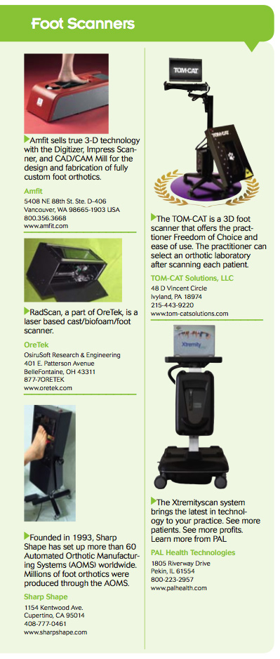
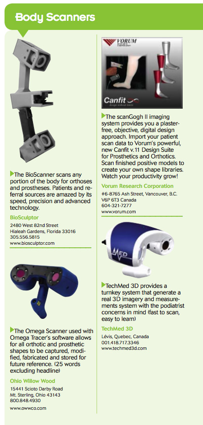
We just reposted the article that includes photos and contact information for the various scanner companies.
Let me know how it all works out.
Regards,
Richard Dubin, Founder and Publisher
Delcam’s iQube is the newest scanner on the market. It meets the criteria discussed in the article.
I am very pleased with this article and I do agree with the author : the 3D optical scanning systems are fare more accurate and user friendly then the other technology in the market.
However, not only is the accuracy of the image that is important for practitioner but also what they do with it.
You want to make sure that you don’t need to spend 20 minutes on the post treatment of the image, you want to have automatic measurements generated from your software and also you want to compare scans automatically in order to trace the evolution of the modification you have applied.
Those benefits are truly tangible.
Marie
Very good article! Please make one correction however. The TomCat scanner is 2D, not 3D.
It is basically a Flat Bed Scanner with software that attempts to calculate an accurate 3D image. Of course, this is not possible as you point out.
Otherwise, thanks for the great information.
Hi dear
our shop needs one feet scanner for making shoe with best measurment.
can you guide me for a smaller & easily function feet scanner.Certainly with low cost.