
 In worker’s compensation cases, including those involving knee osteoarthritis, an expert witness often will be asked to use his or her knowledge of biomechanics to provide an opinion supporting or refuting a causal relationship between work conditions and an overuse injury.
By Steven T. McCaw, PhD
Litigation following personal injury is widespread in today’s society. Determining cause and allocating responsibility are at the heart of financial settlements in both criminal and civil cases. The social and economic consequences of the assigned liability can be enormous. In such cases, particularly those involving workplace injuries, lower extremity biomechanics often plays a key role.
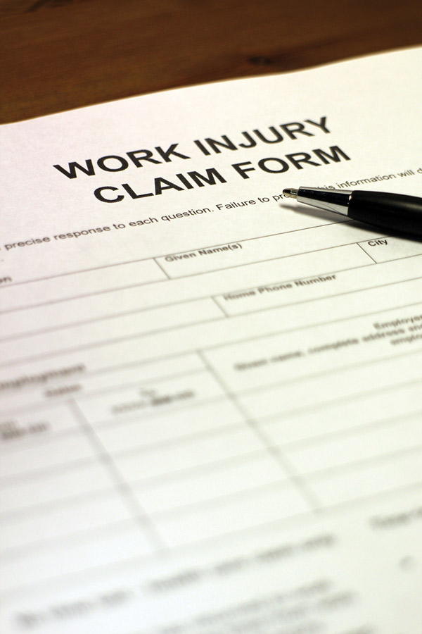
The worker’s compensation (WC) program provides coverage for medical and economic costs, such as lost wages, resulting from a workplace injury. WC programs began in the US in the early 1900s, and since 1949 all states have provided programs.1 Under WC, employees forfeit the right to sue an employer for negligence in exchange for assured, but limited, compensation. WC provides a form of disability, health, and life insurance, with disputed claims adjudicated by a state administrative board overseen by an administrative judge instead of through a trial court. The board resolves contested claims by determining if an injury is work related. Appeals are generally limited, as one purpose of establishing WC was to reduce court adjudication of claims.
Establishing that workplace conditions or events were responsible for an injury can be a contentious issue in WC. In cases of traumatic injury, it’s typically easy to isolate a single causative incident, such as a trip, fall, or crushing event. Overuse injuries are problematic because they develop from repetitive loading and not a single, isolated trauma. The task of separating possible work-related causes from possible away-from-work causes can be contentious in cases involving overuse injury. If the injury did not occur at work, any benefits must be covered through private insurance and not by WC. Claims for overuse injury are frequently challenged by employers, particularly in the manufacturing sector, where WC expenses represent 3% of overall employee costs per hour.2
Unlike the prosecution in a criminal case having to prove an accused individual’s guilt “beyond a reasonable doubt,” in a WC claim, showing causation requires support from a preponderance of the evidence, as carefully woven into a case by an attorney.3 Often, a witness will be asked to use his or her knowledge of biomechanics to provide an expert opinion supporting or refuting a causal relationship between work conditions (identifying a source of the loading) and an overuse injury (developing from a known loading pattern). The opinion must be supported by evidence to a reasonable degree of scientific certainty, with the presiding judge determining the case outcome based on which side best demonstrated that standard.
The Occupational Safety and Health Administration (OSHA) guidelines4 mandate that an employer “shall furnish to each of his employees employment and a place of employment which are free from recognized hazards that are causing or are likely to cause death or serious physical harm.” Many WC disputes on overuse injury arise from the nebulous statements “recognized hazards that are causing or are likely to cause” and “serious harm.”
The biomechanics of osteoarthritis
Osteoarthritis (OA) is a widely prevalent degenerative joint condition, affecting about 12% of the adult population in the US. OA is a “whole joint” disease because every anatomical structure within and around a joint is affected.5 Patterns of OA have been linked with certain occupations,6 and the job-related costs for medical treatment and lost productivity related to OA are in the billions of dollars.7 OA is a form of “serious harm.”
Structurally, knee OA is characterized by the thinning and eventual disappearance of articular cartilage. Damaged cartilage cannot heal because it is nonvascularized. Progressively, the articulating bone surfaces become denser, osteophytes project into the joint space, ligaments loosen, the joint capsule thickens, and surrounding muscles weaken through atrophy and changes in neural drive.8-11 Importantly, the area of the cartilage that provides contact between the bones of the joint becomes smaller as it degenerates, altering the distribution of stress over the joint surfaces.12 In advanced knee OA, the cartilage wears away completely and the surfaces of the now-contacting bones become severely deformed.

Cartilage changes associated with knee OA are typically evident on x-rays or magnetic resonance imaging before symptoms are reported. As mentioned, cartilage is not easily, if at all, repairable. This is important because the joint has typically reached a significant level of deterioration before interventions are started to treat the symptoms and slow progression of disease.
Knee OA interferes with kinesthetic sense, the ability to detect joint position and change of position. Kinesthetic sense is necessary to control the joint and to prevent excessive movement. Loss of position sense leads to additional knee instability,13 and higher joint stress beyond that is caused by mechanical damage to the joint structures. This exacerbates the progression of knee OA and interferes with activities such as walking and stair use.
Clinically, knee OA includes symptoms specific to the joint (pain, perceived weakness, instability, and joint buckling) and symptoms specific to activity (impaired locomotion, ability to work, and recreation). Joint stiffness, swelling, inflammation, tenderness, crepitus, and limited range of motion are typical. Symptoms are aggravated by, and continue following, joint use.
The initiating factors for knee OA remain controversial, and this controversy underlies some companies’ reluctance to pay knee OA-related WC claims. Two hypotheses dominate the discussion of knee OA initiation. The inflammatory hypothesis14 posits that inflammation within the joint capsule initiates the cartilage degeneration. The mechanical hypothesis15 states that joint malalignment or injury alters the stress distribution over the cartilage within the joint to initiate the degenerative process.
What is not controversial is that, regardless of the initiating source, both mechanical loading and inflammation are present as joint failure progresses.14,15 Treatment of the inflammation and other clinical symptoms will not reverse OA, or even slow progression, unless the source of mechanical loading is reduced or eliminated. Continued loading of a degenerating joint makes it inevitable that the degeneration process will continue. The problem is a conundrum in the workplace when repetitive loading is imposed by the design of or the requirements of a task performed at a workstation. For this reason, repetitive loading is a recognized hazard likely to cause serious physical harm in the form of OA.
A case in point
In a disputed WC claim, I was retained as an expert witness for a 57-year-old man following denial of coverage for knee joint replacement. He was morbidly obese with tricompartmental OA in his left knee, and had been employed by a manufacturing company for 36 years. The employer contended that his task, which included stepping up and down a 7-inch step four times per hour, had not caused the OA, and claimed the degeneration resulted from regular activity. The worker countered that several work incidents, including a slip on a bolt four years prior and a severe twisting of his knee when it gave out while he was stepping down from his platform two years earlier, caused knee joint damage that was aggravated by his continued use of the workstation step.
Clinical notes and medical records from physicians are needed to provide a critical foundation for any disability claim related to an overuse injury. Documented symptoms, and the diagnoses, prognoses, treatment plans, and prescriptions, supplemented with notes from physical or occupational therapists, create a timeline for the onset and progression of a condition. In the disputed case, records over the previous five years from the claimant’s personal physician, the company medical team, and an independent orthopedist documented the progression of his OA. This included degeneration of the joint capsule, atrophy of the neuromuscular system, and symptoms including pain, inflammation, perception of instability, impaired movement, and sleep disturbances. The records also summarized the medical interventions that had been used, including diagnostic imaging, arthroscopic surgery (meniscectomy and chondroplasty three years earlier), oral medication, and anti-inflammatory injections. These provided only temporary pain relief and did not slow progression of OA to its advanced stage. There was general consensus that a joint replacement was necessary because conservative treatments had failed.
One aspect of the employer’s liability in the claim was attributed to the design of the workstation, specifically repetitive use of the 7-inch step. The claimant’s lawyer retained my services to provide an opinion on whether the workstation step exacerbated the progression of his OA.
Stair negotiation and knee OA
The three primary functions of the lower extremity during locomotion are energy absorption, energy generation, and support.16 Compared with level walking, each function is more demanding during stair negotiation. During lead foot contact in descent, the knee joint first flexes to absorb the downward momentum, then extends briefly to stabilize the body, and finally flexes again to lower to the next step. The loading of the lower extremity joints is increased by the need for a greater range of motion and higher muscle joint torques when navigating a step.20,21
Difficulty with stair use is a common complaint in patients with knee OA because of increased pain, muscle weakness, and the perception of the knee “giving out.”17 Individuals with knee OA compensate by using a strategy different from healthy individuals during stair use. They use steps more slowly than healthy individuals, which is most evident during stair descent, when controlling the body’s downward momentum is critical.
Ground reaction forces (GRF), the external forces produced at the foot-ground interface to control locomotion, are higher during stair use than during level or sloped walking. The initial impulsive load magnitude at stair contact is about 33% higher and the loading rate is about 68% higher compared with initial contact during walking.18,19 Coordinated joint flexion, controlled by eccentric muscle activity, is important to attenuate these forces as they propagate toward the head. Because they encounter greater absorption and stabilization demands while perceiving joint instability, patients with knee OA typically use a slightly wider step width,18 a more extended knee position at stair contact,18 and less joint flexion during absorption and body lowering than healthy individuals.20 Reconciling the conflict between energy absorption and stability, individuals with knee OA accept higher joint loading to achieve greater joint stability.
 Greater joint torques are required to control individual joint flexion and extension in patients with knee OA than in controls, and these joint torques result from higher levels of muscle activity. The activation level of both the quadriceps and hamstring muscles are slightly higher for individuals with knee OA than for those with healthy knees. This is of great importance because the increased muscle forces around the joint raise the bone-on-bone force within the joint more than can be attributed solely to the fluctuating GRFs. Greater activation of weaker muscles suggests individuals with knee OA use a higher percentage of their functional capacity than healthy individuals.19,21 Thus, individuals with knee OA are more likely than those with healthy knees to fatigue with repetitive step use, further impairing energy absorption and creating a greater risk of falls.21
By descending more slowly and using less knee flexion, individuals with knee OA can reduce their net loading at the knee joint. Because knee OA reduces the joint contact area, however, stress on the remaining cartilage remains high in spite of the reduced loading.22 Even with adaptations, continued step use contributes to the degenerative process.
Criteria for causation
The medical literature is typically based on studies of large samples. But injuries happen to individuals, not populations, and individuals vary in measured response across a sample. To present a convincing expert opinion implicating step use as a contributor to knee OA, it was important to meet three criteria23,24 for causation:
1) Show the OA followed the loading in an appropriate temporal sequence;
2) Present objective evidence of both the loading exposure and the OA development; and
3) Demonstrate the biomechanical plausibility that the loading exposure exceeded the tolerance known to increase risk for knee OA.
These criteria of causation were met by combining the medical records and work history with the literature on the biomechanics of step use. The documented history of reported symptoms, physical evaluations, and medical treatments established the timeline of OA development following the employee’s slip on the bolt. His work history of continued step use without significant modification, other than temporary restrictions immediately following his surgery, constituted repetitive loading that occurred during the period of OA progression. Synthesizing the published literature on knee loading during step use and on knee OA pathogenesis demonstrated the biomechanical plausibility of step use at work being a primary contributor to onset and progression of the employee’s knee OA.
The decision
With these criteria covered, it was not difficult to confidently provide and defend an opinion that the step use at work was a probable causative factor in the onset and progression of OA necessitating knee joint replacement. The administrative board accepted this as a convincing opinion and ruled that the employee’s OA was work-related. However, because of the possible precedent-setting nature of the decision as it applies to the widespread prevalence of OA in workers, the case is under appeal.
Steven T. McCaw, PhD, is a professor emeritus in exercise science from Illinois State University in Normal and now consults on personal injury cases through McCaw Biomechanics Consulting in Charleston, IL.
- Guyton GP. A brief history of worker’s compensation. Iowa Orthop J 1999;19:106-110.
- National Compensation Survey. Table 5. Employer costs per hour worked for employee compensation and costs as a percent of total compensation: Private industry workers, by major occupational group and bargaining unit status. Bureau of Labor Statistics. US Department of Labor website. http://www.bls.gov/news.release/ecec.t05.htm. Updated March 10, 2016. Accessed June 3, 2016.
- Schneck J. Forensic biomechanics. Am Lab News 2005;37(8):4-5.
- OSHA Act of 1970. Occupational Safety & Health Administration website. https://www.osha.gov/pls/oshaweb/owadisp.show_document?p_table=OSHACT&p_id=2743. Published December 29, 1970. Amended January 1, 2004. Accessed June 3, 2016.
- Radin EL. Who gets osteoarthritis and why? J Rheum Suppl 2004;70:10-15.
- Yucesoy B, Charles LE, Baker B, Burchfiel CM. Occupational and genetic risk factors for osteoarthritis: A review. Work 2015;50(2):261-273.
- Yelin E, Murphy L, Cisternas M, et al. Medical care expenditures and earnings losses among persons with arthritis and other rheumatic conditions in 2003, and comparisons to 1997. Arthritis Rheum 2007;56(5):1397-1407.
- Bennell KL, Hinman RS, Metcalf BR. Association of sensorimotor function with knee joint kinematics during locomotion in knee osteoarthritis. Am J Phys Med Rehabil 2004;83(6):455-463.
- Fitzgerald GK, Piva SR, Irrgang JJ. Reports of joint instability in knee osteoarthritis: its prevalence and relationship to physical function. Arthritis Rheum 2004;51(6):941-946.
- Roos EM, Herzog W, Block JA, Bennell KL. Muscle weakness, afferent sensory dysfunction and exercise in knee osteoarthritis. Nat Rev Rheumatol 2011;7(1):57-63.
- Ageberg E, Roos EM. Neuromuscular exercise as treatment of degenerative knee disease. Exerc Sport Sci Rev 2015;43(1):14-22.
- Tummala S, Nielsen M, Lillholm M, et al. Automatic quantification of tibio-femoral contact area and congruity. IEEE Trans Med Imag 2012;31(7):1404-1412.
- Blalock D, Miller A, Tilley M, Wang J. Joint instability and osteoarthritis. Clin Med Insights Arthritis Musculoskelet Disord 2015;8:15-23.
- Berenbaum F. Osteoarthritis as an inflammatory disease (osteoarthritis is not osteoarthrosis!). Osteoarthritis Cartilage 2013;21(1):16-21.
- Felson DT. Osteoarthritis as a disease of mechanics. Osteoarthritis Cartilage 2013;21(1):10-15.
- Winter DA, Eng P. Kinetics: our window into the goals and strategies of the central nervous system. Behav Brain Res 1995;67(2):111-120.
- Nguyen US, Felson DT, Niu J, et al. The impact of knee instability with and without buckling on balance confidence, fear of falling and physical function: The Multicenter Osteoarthritis Study. Osteoarthritis Cartilage 2014;22(4):527–534.
- Liikavainio T, Isolehto J, Helminen HJ, et al. Loading and gait symmetry during level and stair walking in asymptomatic subjects with knee osteoarthritis: Importance of quadriceps femoris in reducing impact force during heel strike. Knee 2007;14(3):231-238.
- Liikavainio T, Bragge T, Hakkarainen M, et al. Gait and muscle activation changes in men with knee osteoarthritis. Knee 2010;17(1):69-76.
- Hicks-Little CA, Peindl RD, Fehring TK, et al. Temporal-spatial gait adaptations during stair ascent and descent in patients with knee osteoarthritis. J Arthroplasty 2012;27(6):1183-1187.
- Novak AC, Brouwer B. Sagittal and frontal lower limb joint moments during stair ascent and descent in young and older adults. Gait Posture 2011;33(1):54-60.
- Adouni M, Shirazi-Adl A. Evaluation of knee joint muscle forces and tissue stresses-strains during gait in severe OA versus normal subjects. J Orthop Res 2014;32(1):69-78.
- Hill AB. The environment and disease: association or causation? Proc R Soc Med 1965;58(5):295-300.
- Hayes WC, Erickson MS, Power ED. Forensic injury biomechanics. Annu Rev Biomed Eng 2007;9:55-86.


